Detection of Cerebral Atrophy and Vascular Disorders in Patients with Mature Extra-cerebral Tumours in Pre- and Postoperative Period According to Morphometric Analysis of Magnetic Resonance Tomography Findings
The aim of the investigation is to study morphological background of neurological disorders in patients with extracerebral intracranial tumours in pre- and postoperative period (after 1 and 2 years after the operation) using morphometric analysis.
Materials and Methods. 50 patients with extracerebral intracranial tumours (31 patients with supratentorial tumours, 19 — with infratentorial tumours) before the operation, and 1 and 2 years after the operation were studied. The patients underwent clinico-neurological examination, brain MRT with measurement of lateral ventricles width, subarachnoid space, and vascular foci. The control group consisted of 30 people without focal cerebral lesions according to MRT findings.
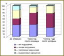
Results. The processes of mixed cerebral atrophy both in supratentorial and infratentorial tumours were stated to become more intense after the operation. The morphometric analysis confirmed the progress of the stated changes. The second revealed characteristic of MRT-picture in this group of patients in postoperative period was vascular foci formation in white matter.
- Gusev E.I., Konovalov A.N., Burd G.S. Nevrologiya i neyrokhirurgiya [Neurology and neurosurgery]. Moscow: Meditsina; 2000; 656 p.
- Gunderson L.L., Tepper J.E. Clinical radiation oncology. Edinbourgh; 2006.
- Handbook of evidence-based radiation oncology. E.K. Hansen, M. Roach (editors). London; 2007.
- Avoyan K.M., Muratova S.M. Med Pomos? 2006; 3: 8–10.
- Meller T.B., Rayf E. Norma pri KT i MRT issledovaniyah [Norm in CT and MRT studies]. Pod red. G.E. Trufanova, N.V. Marchenko [G.E. Trufanova, N.V. Marchenko (editors)]. Moscow; 2008.
- Yakhno N.N., Levin O.S., Damulin I.V. Nevrol Z 2001; 3: 10–19.


