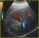
Normal Echosemiotics of Resected Parenchymatous Organs
The aim of the investigation was to develop ultrasonic techniques of the resected liver, pancreas, and kidney, and study their normal echosemiotics after different types of resection.
Materials and Methods. We examined 404 patients after various hepatectomies, 145 — after extended pancreaticoduodenal resections, and 123 — after different types of nephrectomy. Ultrasound was performed on scanners Voluson 730 PRO (GE, USA) and Technos (Esaote, Italy) in early postoperative period — on day 2–3 and on day 7–10, and in follow-up care — 3, 6 and 12 months after the operation.
Results. We developed an ultrasound technique and established sonographic criteria to assess resected parenchymatous organs, represented normal ultrasound semiotics of the liver, pancreas and kidney after different types of resection. The number and location of hepatic veins in hepatic stump was found to be of primary importance in determining the hepatectomy type; and it was called the hepatic vein rule. An additional criterion was the portal vein branching character. The assessment criteria of pancreatic stump were its size in the body of pancreas and diameter of the major pancreatic duct, as well as spatial location of anastomosed loop of jejunum and gastric remnant. During the first postoperative month slight dilatation of Wirsung duct up to 3–4 mm with its following recovery is permissible. In late postoperative period, the duct dilatation over 3 mm is considered pathological. To determine the nephrectomy type it is necessary to assess the form of the organ and the resection area. Normal echogram can be misinterpreted after frontal nephrectomy due to different parenchymal thickness in its resected and remaining parts.
Conclusion. The developed echosemiotics of resected parenchymatous organ in the majority of cases enables to take a correct view of the volume and character of the surgery, and determine postoperative state of the stump.
- Prakticheskoe rukovodstvo po ul’trazvukovoy diagnostike. Obshchaya ul'trazvukovaya diagnostika [Practice guidelines on diagnostic ultrasound. General diagnostic ultrasound]. Pod red. Mit’kova V.V. [Mit’kov V.V. (editor)]. Moscow: Izdatel’skiy dom Vidar-M; 2003; 720 p.
- Wermke W. Sonographische differenzialdiagnose leberkrankheiten. Lehrbuch und systematischer Atlas [Sonographic differential diagnostics of liver diseases. Manual and systematic atlas]. Köln: Deutscher rzte-Verlag; 2006; 446 p.
- Vishnevskiy V.A., Efanov M.G., Ikramov R.Z. Prakticheskie aspekty sovremennoy khirurgii pecheni [Practical aspects of modern hepatic surgery]. Tikhookeanskiy meditsinskiy zhurnal — Pacific Ocean Medical Journal 2009; 2: 28–34.
- Atduev V.A., Ovchinnikov V.A. Khirurgiya opukholey parenkhimy pochki [Surgery of renal parenchyma tumors]. Moscow: Med. kniga; 2004; 191 p.
- Patyutko Yu.I. Khirurgicheskoe lechenie zlokachestvennykh opukholey pecheni [Surgical treatment of hepatic tumors]. Moscow: Prakticheskaya meditsina; 2005; 312 p.
- Alyaev Yu.G., Krapivin A.A. Rezektsiya pochki pri rake [Partial nephrectomy in cancer]. Moscow: Meditsina; 2001; 224 p.
- Patyutko Yu.I., Kotel‘nikov A.G. Khirurgiya raka organov biliopankreatoduodenal‘noy zony [Surgery of biliopancreatoduodenal tumors]. Moscow: Meditsina; 2007; 448 p.
- Karmazanovskiy G.G., Fedorov V.D., Shipuleva I.V. Spiral‘naya komp‘yuternaya tomografiya v khirurgicheskoy gepatologii [Helical computer tomography in surgical hepatology]. Moscow: Russkiy vrach; 2000; 150 p.


