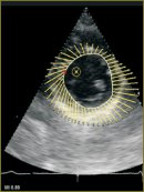
VVI Technique Possibilities in the Assessment of the Indices of Left Ventricular Systolic Function and All Its Segments
The aim of the investigation was to estimate Velocity Vector Imaging (VVI) feasibility in the study of left ventricular (LV) and all its segments of systolic function in healthy volunteers when developing standards.
Materials and Methods. 26 healthy volunteers without cardiovascular pathology were recruited and participated in the survey, their mean age being 21.7±3.0 years. LV systolic function using VVI was studied in apical 4-, 5- and 2-chamber views and in parasternal view along a short axis at the level of the mitral valve (LV basal segments), papillary muscles (LV middle segments), and the apex (LV apical segments). We analyzed the following indices: myocardial movement rate, strain (deformation), strain rate (rate of deformation), LV ejection fraction and volume.
Results. The comparison of LV systolic function indices using standard echocardiography and VVI technique showed VVI value in the assessment of the parameters to be rather high and able to acquire objective data on a large number of parameters. VVI enables to record even minimal LV dysfunctions, and the analysis of longitudinal, radial and circular fibers enables to assess transmural damage and reveal LV dysfunction mechanism. The indices of longitudinal, circular and radial deformation averaged –19.9±2.6, –21.6±5.5 and 32.3±7.6%, respectively. Strain rate of longitudinal, circular and radial fibers were –1.17±0.26, –1.32±0.44 and 1.58±0.32 s–1, respectively. LV systolic function indices obtained using VVI can serve as a norm in the assessment of the functioning of LV and all its segments.
- Funktsional’naya diagnostika v kardiologii: klinicheskaya interpretatsiya [Functional diagnostics in cardiology: clinical interpretation]. Pod red. Vasyuka Yu.A. [VasyukaYu.A. (editor)]. Moscow: Prakticheskaya meditsina; 2009; 312 p.
- Tkachenko S.B., Beresten’ N.F. Tkanevoe doplerovskoe issledovanie miokarda [Doppler tissue myocardial technique]. Moscow: “Real Time”; 2006; 176 p.
- Kostis J.B., Mavrogeorgis E., Slater A., et al. Use of range-gated, pulsed ultrasonic Doppler technique for continuous measurement of velocity of the posterior heart wall. Chest 1972; 62: 597–604.
- Sonnenblick E.H., Parmley W.W., Urschel C.W., Brutsaert D.L. Ventricular function: evaluation of myocardial contractility in health and disease. Prog Cardiovasc Dis 1970; 12: 449–466.
- Mirsky I., Parmley W.W. Assessment of passive elastic stiffness for isolated heart muscle and the intact heart. Circ Res 1973; 33: 233–243.
- Isaaz K., Thompson A., Ethevenot G., et al. Doppler echocardiographic measurement of low velocity motion of the left ventricular posterior wall. Am J Cardiol 1989; 64: 66–75.
- Sutherland G.R., Stewart M.J., Groundstroem K.W., et al. Color Doppler myocardial imaging: a new technique for the assessment of myocardial function. J Am Soc Echocardiogr 1994; 7: 441–458.
- Yamazaki N., Mine Y., Sano A., et al. Analysis of ventricular wall motion using color — coded tissue Doppler imaging system. Jpn J Appl Rhys 1994; 33: 3141–3146.
- Miyatake K., Yamagishi M., Tanaka N., et al. New method for evaluating left ventricular wall motion by color — coded tissue Doppler imaging: in vitro and in vivo studies. J Am Coll Cardiol 1995; 25: 717–724.
- Fleming A.D., Xia X., McDicken W.N., et al. Myocardial velocity gradients detected by Doppler imaging. Br J Radiol 1994; 67: 679–688.
- Heimdal A., Dhogge J., Bijnens B., et al. In vitro validation of in-plane strain rate imaging. A new ultrasound technique for evaluating regional myocardial deformation based on tissue Doppler imaging. Echocardiography 1998; 15(8-II): S 40.
- Nikitin N.P., Kliland D.D. Primenenie tkanevoy miokardial’noy doppler-ekhokardiografii v kardiologii [The use of tissue myocardial Doppler echocardiography in cardiology]. Kardiologiya — Cardiology 2002; 3: 66–79.
- Carasso Sh., Biaggi P., Rakowski H., et al. Velocity vector imaging: standard tissue-tracking results acquired in normals — the VVI-strain study. Am Soc Echocardiography 2012; 25(5): 543–552.
- Mani A. Vannan., Gianni Pedrizzetti, Peng Li., et al. Effect of cardiac resynchronization therapy on longitudinal and circumferential left ventricular mechanics by velocity vector imaging: description and initial clinical application of a novel method using high-frame rate B-mode echocardiographic images. Ecocardiography 2005; 10: 826–830.
- Syvolap V.V., Kolesnik M.Yu. Otsenka prodol’noy i radial’noy sistolicheskoy deformatsii miokarda levogo zheludochka pri dilatatsionnoy kardiomiopatii (klinicheskoe nablyudenie) [The assessment of longitudinal and radial systolic deformation of the left ventricular myocardium in dilated cardiomyopathy (clinical observation)]. Vnutrennyaya meditsina — Internal Medicine 2008; 5–6(11–12).
- Voight J.U., Flachskampf F.A. Strain and strain rate. New and clinically relevant echo parameters of regional myocardial function. Z Kardiol 2004; 93: 249–258.
- Alekhin M.N. Vozmozhnosti prakticheskogo ispol’zovaniya tkanevogo dopplera. Lektsiya 1. Tkanevoy doppler, printsipy raboty i ego osobennosti [The possibilities of tissue Doppler practical application. Lecture 1. Tissue Doppler, principles of functioning and its characteristics]. Ul’trazvukovaya i funktsional’naya diagnostika — Ultrasound and Functional Diagnosis 2002; 3: 90–98.
- Kowalski M., Kukulski T., Jamal F., et al. Can natural strain and strain rate quantify regional myocardial deformation? A study in healthy subjects. Ultrasound Med Biol 2001; 27: 1087–1097.
- Garot J., Derumeaux G.A., Monin J.L., et al. Quantitative systolic and diastolic transmyocardial velocity gradients assessed by M-mode colour Doppler tissue imaging as reliable indicators of regional left ventricular function after myocardial infarction. Eur Heart J 1999 Apr; 20(8): 593–603.
- Alekhin M.N. Vozmozhnosti prakticheskogo ispol’zovaniya tkanevogo dopplera. Lektsiya 2. Tkanevoy doppler fibroznykh kolets atrioventrikulyarnykh klapanov [The possibilities of tissue Doppler practical application. Lecture 2. Tissue Doppler imaging of fibrous rings of atrioventricular valves]. Ul’trazvukovaya i funktsional’naya diagnostika — Ultrasound and Functional Diagnosis 2002; 4: 112–118.
- Notomi Y., Srinath G., Shiota T., et al. Maturational and adaptive modulation of left ventricular torsional biomechanics: Doppler tissue imaging observation from infancy to adulthood. Circulation 2006; 113(21): 2534–2541.


