Multifunctional 1050 nm Spectral Domain OCT System at 147 kHz for Posterior Eye Imaging
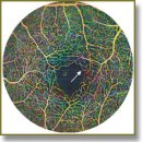
The recent development of optical coherence tomography in ophthalmology has shown great interests in using the system in 1 µm in contrast to 800 nm wavelength range due to the less reflection and absorption of retinal pigment epithelium and pigmented choroidal melanocytes in 1 µm wavelength. The clinical value of using 1 µm system has been demonstrated in choroid imaging, retinal and choroidal microcirculation, etc. By examining different aspects of the posterior eye, the specificity and sensitivity of diagnosis can be increased. On the other hand, higher speed can greatly reduce the measuring time and motion artifacts, which brings comfort to the patients and improves the image quality. In this work, we report a newly developed multifunctional 1050 nm spectral domain optical coherence tomography (SD-OCT) system working at 147 kHz A-scan rate for posterior eye imaging. The uniqueness of this system is: 1) its capability of providing not only simultaneous structural imaging of the complete posterior eye, but also the visualization of the retinal blood vessel network with larger field of view and good image quality compared with former SD-OCT systems; 2) it’s fast 147 kHz A-scan rate which has not been reported before. It is demonstrated through in vivo experiments that this system delivers not only superior performance of posterior eye structural imaging but also detailed visualization of microcirculation network in retina. The choroid of the eye with either myopic or normal conditions can clearly be visualized through the entire scanning volume. These results indicate great potential in applying this new system for clinical studies.
- Fujimoto J.G., Pitris C., Boppart S.A., Brezinski M.E. Optical coherence tomography: an emerging technology for biomedical imaging and optical biopsy. Neoplasia 2000; 2(1–2): 9–25, http://dx.doi.org/10.1038/sj.neo.7900071.
- Hee M.R., Puliafito C.A., Duker J.S., Reichel E., Coker J.G., Wilkins J.R., Schuman J.S., Swanson E.A., Fujimoto J.G. Topography of diabetic macular edema with optical coherence tomography. Ophthalmology 1998; 105(2): 360–370, http://dx.doi.org/10.1016/s0161-6420(98)93601-6.
- Esmaeelpour M., Ansari-Shahrezaei S., Glittenberg C., Nemetz S., Kraus M.F., Hornegger J., Fujimoto J.G., Drexler W., Binder S. Choroid, Haller’s, and Sattler’s layer thickness in intermediate age-related macular degeneration with and without fellow neovascular eyes. Invest Ophthalmol Vis Sci 2014; 55(8): 5074–5080, http://dx.doi.org/10.1167/iovs.14-14646.
- An L., Qin J., Wang R.K. Ultrahigh sensitive optical microangiography for in vivo imaging of microcirculations within human skin tissue beds. Opt Express 2010; 18(8): 8220–8228, http://dx.doi.org/10.1364/OE.18.008220.
- Fernández E.J., Hermann B., Považay B., Unterhuber A., Sattmann H., Hofer B., Ahnelt P.K., Drexler W. Ultrahigh resolution optical coherence tomography and pancorrection for cellular imaging of the living human retina. Opt Express 2008; 16(15): 11083–11094, http://dx.doi.org/10.1364/OE.16.011083.
- Flores-Moreno I., Lugo F., Duker J.S., Ruiz-Moreno J.M. The relationship between axial length and choroidal thickness in eyes with high myopia. Am J Ophthalmol 2013; 155(2): 314–319.e1, http://dx.doi.org/10.1016/j.ajo.2012.07.015.
- Klein T., Wieser W., Eigenwillig C.M., Biedermann B.R., Huber R. Megahertz OCT for ultrawide-field retinal imaging with a 1050 nm Fourier domain mode-locked laser. Opt Express 2011; 19(4): 3044–3062, http://dx.doi.org/10.1364/OE.19.003044.
- Choi W., Mohler K.J., Potsaid B., Lu C.D., Liu J.J., Jayaraman V., Cable A.E., Duker J.S., Huber R., Fujimoto J.G. Choriocapillaris and choroidal microvasculature imaging with ultrahigh speed OCT angiography. PLoS ONE 2013; 8(12): e81499, http://dx.doi.org/10.1371/journal.pone.0081499.
- Haas P., Esmaeelpour M., Ansari-Shahrezaei S., Drexler W., Binder S. Choroidal thickness in patients with reticular pseudodrusen using 3D 1060-nm OCT maps. Invest Ophthalmol Vis Sci 2014; 55(4): 2674–2681, http://dx.doi.org/10.1167/iovs.13-13338.
- Považay B., Hermann B., Hofer B., Kajić V., Simpson E., Bridgford T., Drexler W. Wide-field optical coherence tomography of the choroid in vivo. Invest Ophthalmol Vis Sci 2009; 50(4): 1856–1863, http://dx.doi.org/10.1167/iovs.08-2869.
- Wang R.K., An L. Multifunctional imaging of human retina and choroid with 1050-nm spectral domain optical coherence tomography at 92-kHz line scan rate. J Biomed Opt 2011; 16(5): 050503, http://dx.doi.org/10.1117/1.3582159.
- An L., Li P., Lan G., Malchow D., Wang R.K. High-resolution 1050 nm spectral domain retinal optical coherence tomography at 120 kHz A-scan rate with 6.1 mm imaging depth. Opt Express 2013; 4(2): 245–259, http://dx.doi.org/10.1364/BOE.4.000245.
- American National Standard Institute. Safe use of lasers and safe use of optical fiber communications. New York: ANSI; 2000; No.168.
- Flores-Moreno I., Lugo F., Duker J.S., Ruiz-Moreno J.M. The relationship between axial length and choroidal thickness in eyes with high myopia. Am J Ophthalmol 2013; 155(2): 314–319, http://dx.doi.org/10.1016/j.ajo.2012.07.015.
- Yin X., Chao J.R., Wang R.K. User-guided segmentation for volumetric retinal optical coherence tomography images. J Biomed Opt 2014; 19(8): 086020, http://dx.doi.org/10.1117/1.JBO.19.8.086020.
- An L., Shen T.T., Wang R.K. Using ultrahigh sensitive optical microangiography to achieve comprehensive depth resolved microvasculature mapping for human retina. J Biomed Opt 2011; 16(10): 106013, http://dx.doi.org/10.1117/1.3642638.


