Detection of Atherosclerotic Plaque from Optical Coherence Tomography Images Using Texture-Based Segmentation
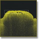
Detection of atherosclerotic plaque from optical coherence tomography (OCT) images by visual inspection is difficult. We developed a texture based segmentation method to identify atherosclerotic plaque automatically from OCT images without any reliance on visual inspection. Our method involves extraction of texture statistical features (spatial gray level dependence matrix method), application of an unsupervised clustering algorithm (K-means) on these features, and mapping of the clustered regions: background, plaque, vascular tissue and an OCT degraded signal region in feature-space, back to the actual image. We verified the validity of our results by visual comparison to photographs of the vascular tissue with atherosclerotic plaque that were used to generate our OCT images. Our method could be potentially used in clinical studies in OCT imaging of atherosclerotic plaque.
- Hoyert D.L., Xu J. Deaths: preliminary data for 2011. Nat Vital Stat Rep 2012; 61(6): 1–51.
- Lloyd-Jones D., Adams R.J., Brown T.M., Carnethon M., Dai S., De Simone G., Ferguson T.B., Ford E., Furie K., Gillespie C., Go A., Greenlund K., Haase N., Hailpern S., Ho P.M., Howard V., Kissela B., Kittner S., Lackland D., Lisabeth L., Marelli A., McDermott M.M., Meigs J., Mozaffarian D., Mussolino M., Nichol G., Roger V.L., Rosamond W., Sacco R., Sorlie P., Stafford R., Thom T., Wasserthiel-Smoller S., Wong N.D., Wylie-Rosett J. Heart disease and stroke statistics — 2010 update a report from the American Heart Association. Circulation 2010; 121(7): e46–e215, http://dx.doi.org/10.1161/circulationaha.109.192667.
- Burke A.P., Farb A., Malcom G.T., Liang Y., Smialek J., Virmani R. Coronary risk factors and plaque morphology in men with coronary disease who died suddenly. N Engl J Med 1997; 336(18): 1276–1282, http://dx.doi.org/10.1056/NEJM199705013361802.
- Fuster V., Moreno P.R., Fayad Z.A., Corti R., Badimon J.J. Atherothrombosis and high-risk plaque: part I: evolving concepts. J Am Coll Cardiol 2005; 46(6): 937–954, http://dx.doi.org/10.1016/j.jacc.2005.03.074.
- Takayama T., Hiro T., Yamagishi M., Daida H., Hirayama A., Saito S., Yamaguchi T., Matsuzaki M.; The COSMOS investigators. Effect of rosuvastatin on coronary atheroma in stable coronary artery disease: multicenter coronary atherosclerosis study measuring effects of rosuvastatin using intravascular ultrasound in Japanese subjects (COSMOS). Cir J 2009; 73(11): 2110–2117, http://dx.doi.org/10.1253/circj.CJ-09-0358.
- Kubo T., Maehara A., Mintz G.S., Doi H., Tsujita K., Choi S.-Y., Katoh O., Nasu K., Koenig A., Pieper M., Rogers J.H., Wijns W., Böse D., Margolis M.P., Moses J.W., Stone G.W., Leon M.B. The dynamic nature of coronary artery lesion morphology assessed by serial virtual histology intravascular ultrasound tissue characterization. J Am Coll Cardiol 2010; 55(15): 1590–1597, http://dx.doi.org/10.1016/j.jacc.2009.07.078.
- Lee C.-H., Tai B.-C., Soon C.-Y., Low A.F., Poh K.-K., Yeo T.-C., Lim G.-H., Yip J., Omar A.R., Teo S.-G., Tan H.-C. New set of intravascular ultrasound-derived anatomic criteria for defining functionally significant stenoses in small coronary arteries (results from intravascular ultrasound diagnostic evaluation of atherosclerosis in Singapore [IDEAS] study). Am J Cardiol 2010; 105(10): 1378–1384, http://dx.doi.org/10.1016/j.amjcard.2010.01.002.
- Arbab-Zadeh A., Miller J.M., Rochitte C.E., Dewey M., Niinuma H., Gottlieb I., Paul N., Clouse M.E., Shapiro E.P., Hoe J., Lardo A.C., Bush D.E., de Roos A., Cox C., Brinker J., Lima J.A.C. Diagnostic accuracy of computed tomography coronary angiography according to pre-test probability of coronary artery disease and severity of coronary arterial calcification: the CORE-64 (coronary artery evaluation using 64-row multidetector computed tomography angiography) international multicenter study. J Am Coll Cardiol 2012; 59(4): 379–387, http://dx.doi.org/10.1016/j.jacc.2011.06.079.
- de Graaf F.R., Schuijf J.D., van Velzen J.E., Kroft L.J., de Roos A., Reiber J.H.C., Boersma E., Schalij M.J., Spanó F., Jukema J.W., van der Wall E.E., Bax J.J. Diagnostic accuracy of 320-row multidetector computed tomography coronary angiography in the non-invasive evaluation of significant coronary artery disease. Eur Heart J 2010; 31(15): 1908–1915, http://dx.doi.org/10.1093/eurheartj/ehp571.
- Bamberg F., Sommer W.H., Hoffmann V., Achenbach S., Nikolaou K., Conen D., Reiser M.F., Hoffmann U., Becker C.R. Meta-analysis and systematic review of the long-term predictive value of assessment of coronary atherosclerosis by contrast-enhanced coronary computed tomography angiography. J Am Coll Cardiol 2011; 57(24): 2426–2436, http://dx.doi.org/10.1016/j.jacc.2010.12.043.
- Underhill H.R., Hatsukami T.S., Fayad Z.A., Fuster V., Yuan C. MRI of carotid atherosclerosis: clinical implications and future directions. Nat Rev Cardiol 2010; 7(3): 165–173, http://dx.doi.org/10.1038/nrcardio.2009.246.
- Kwee R.M., van Oostenbrugge R.J., Mess W.H., Prins M.H., van der Geest R.J., ter Berg J.W.M., Franke C.L., Korten A.G.G.C., Meems B.J., van Engelshoven J.M.A., Wildberger J.E., Kooi M.E. MRI of carotid atherosclerosis to identify TIA and stroke patients who are at risk of a recurrence. J Magn Reson Imaging 2013; 37(5): 1189–1194, http://dx.doi.org/10.1002/jmri.23918.
- Bezerra H.G., Attizzani G.F., Sirbu V., Musumeci G., Lortkipanidze N., Fujino Y., Wang W., Nakamura S., Erglis A., Guagliumi G., Costa M.A. Optical coherence tomography versus intravascular ultrasound to evaluate coronary artery disease and percutaneous coronary intervention. JACC Cardiovasc Interv 2013; 6(3): 228–236, http://dx.doi.org/10.1016/j.jcin.2012.09.017.
- Parodi G., Maehara A., Giuliani G., Kubo T., Mintz G.S., Migliorini A., Valenti R., Carrabba N., Antoniucci D. Optical coherence tomography in unprotected left main coronary artery stenting. EuroIntervention 2010; 6(1): 94–99, http://dx.doi.org/10.4244/eijv6i1a14.
- Foin N., Mari J.M., Davies J.E., Di Mario C., Girard M.J.A. Imaging of coronary artery plaques using contrast-enhanced optical coherence tomography. Eur Heart J Cardiovasc Imaging 2013; 14(1): 85, http://dx.doi.org/10.1093/ehjci/jes151.
- Wang T., Wieser W., Springeling G., Beurskens R., Lancee C.T., Pfeiffer T., van der Steen A.F.W., Huber R., van Soest G. Intravascular optical coherence tomography imaging at 3200 frames per second. Opt Lett 2013; 38(10): 1715–1717, http://dx.doi.org/10.1364/OL.38.001715.
- Gubarkova E.V., Kirillin M.Yu., Sergeeva E.A., Kiseleva E.B., Snopova L.B., Prodanets N.N., Sharabrin L.G., Shakhov E.B., Nemirova S.V., Gladkova N.D. Cross-polarization optical coherence tomography in evaluation of atherosclerotic plaque structure. Sovremennye tehnologii v medicine 2013; 5(4): 45–54.
- van Soest G., Goderie T., Regar E., Koljenović S., van Leenders G.L.J.H., Gonzalo N., van Noorden S., Okamura T., Bouma B.E., Tearney G.J., Oosterhuis J.W., Serruys P.W., van der Steen A.F. Atherosclerotic tissue characterization in vivo by optical coherence tomography attenuation imaging. J Biomed Opt 2010; 15(1): 011105, http://dx.doi.org/10.1117/1.3280271.
- Prati F., Regar E., Mintz G.S., Arbustini E., Di Mario C., Jang I.-K., Akasaka T., Costa M., Guagliumi G., Grube E., Ozaki Y., Pinto F., Serruys P.W.J. Expert review document on methodology, terminology, and clinical applications of optical coherence tomography: physical principles, methodology of image acquisition, and clinical application for assessment of coronary arteries and atherosclerosis. Eur Heart J 2010; 31(4): 401–415, http://dx.doi.org/10.1093/eurheartj/ehp433.
- Li Q.-X., Fu Q.-Q., Shi S.-W., Wang Y.-F., Xie J.-J., Yu X., Cheng X., Liao Y.-H. Relationship between plasma inflammatory markers and plaque fibrous cap thickness determined by intravascular optical coherence tomography. Heart 2010; 96(3): 196–201, http://dx.doi.org/10.1136/hrt.2009.175455.
- Brezinski M.E., Tearney G.J., Bouma B.E., Boppart S.A., Hee M.R., Swanson E.A., Southern J.F., Fujimoto J.G. Imaging of coronary artery microstructure (in vitro) with optical coherence tomography. Am J Cardiol 1996; 77(1): 92–93, http://dx.doi.org/10.1016/S0002-9149(97)89143-6.
- Gossage K.W., Tkaczyk T.S., Rodriguez J.J., Barton J.K. Texture analysis of optical coherence tomography images: feasibility for tissue classification. J Biomed Opt 2003; 8(3): 570–575, http://dx.doi.org/10.1117/1.1577575.
- Lindenmaier A.A., Conroy L., Farhat G., DaCosta R.S., Flueraru C., Vitkin I.A. Texture analysis of optical coherence tomography speckle for characterizing biological tissues in vivo. Opt Lett 2013; 38(8): 1280–1282, http://dx.doi.org/10.1364/OL.38.001280.
- Quellec G., Lee K., Dolejsi M., Garvin M.K., Abràmoff M.D., Sonka M. Three-dimensional analysis of retinal layer texture: identification of fluid-filled regions in SD-OCT of the macula. IEEE Trans Med Imaging 2010; 29(6): 1321–1330, http://dx.doi.org/10.1109/TMI.2010.2047023.
- Liu L., Gardecki J.A., Nadkarni S.K., Toussaint J.D., Yagi Y., Bouma B.E., Tearney G.J. Imaging the subcellular structure of human coronary atherosclerosis using micro-optical coherence tomography. Nat Med 2011; 17(8): 1010–1014, http://dx.doi.org/10.1038/nm.2409.
- Takano M., Yamamoto M., Inami S., Murakami D., Ohba T., Seino Y., Mizuno K. Appearance of lipid-laden intima and neovascularization after implantation of bare-metal stents extended late-phase observation by intracoronary optical coherence tomography. J Am Coll Cardiol 2009; 55(1): 26–32, http://dx.doi.org/10.1016/j.jacc.2009.08.032.
- Mizukoshi M., Imanishi T., Tanaka A., Kubo T., Liu Y., Takarada S., Kitabata H., Tanimoto T., Komukai K., Ishibashi K., Akasaka T. Clinical classification and plaque morphology determined by optical coherence tomography in unstable angina pectoris. Am J Cardiol 2010; 106(3): 323–328, http://dx.doi.org/10.1016/j.amjcard.2010.03.027.
- Uemura S., Ishigami K., Soeda T., Okayama S., Sung J.H., Nakagawa H., Somekawa S., Takeda Y., Kawata H., Horii M., Saito Y. Thin-cap fibroatheroma and microchannel findings in optical coherence tomography correlate with subsequent progression of coronary atheromatous plaques. Eur Heart J 2012; 33(1): 78–85, http://dx.doi.org/10.1093/eurheartj/ehr284.
- Hoogi A., Adam D., Hoffman A., Kerner H., Reisner S., Gaitini D. Carotid plaque vulnerability: quantification of neovascularization on contrast-enhanced ultrasound with histopathologic correlation. AJR Am J Roentgenol 2011; 196(2): 431–436, http://dx.doi.org/10.2214/AJR.10.4522.
- Partovi S., Loebe M., Aschwanden M., Baldi T., Jäger K.A., Feinstein S.B., Staub D. Contrast-enhanced ultrasound for assessing carotid atherosclerotic plaque lesions. AJR Am J Roentgenol 2012; 198(1): W13–W19, http://dx.doi.org/10.2214/AJR.11.7312.
- Vukadinovic D., Rozie S., van Gils M., van Walsum T., Manniesing R., van der Lugt A., Niessen W.J. Automated versus manual segmentation of atherosclerotic carotid plaque volume and components in CTA: associations with cardiovascular risk factors. Int J Cardiovasc Imaging 2012; 28(4): 877–887, http://dx.doi.org/10.1007/s10554-011-9890-6.
- Morishige K., Kacher D.F., Libby P., Josephson L., Ganz P., Weissleder R., Aikawa M. High-resolution magnetic resonance imaging enhanced with superparamagnetic nanoparticles measures macrophage burden in atherosclerosis. Circulation 2010; 122(17): 1707–1715, http://dx.doi.org/10.1161/CIRCULATIONAHA.109.891804.
- Jang I.-K., Bouma B.E., Kang D.-H., Park S.-J., Park S.-W., Seung K.-B., Choi K.-B., Shishkov M., Schlendorf K., Pomerantsev E., Houser S.L., Aretz H.T., Tearney G.J. Visualization of coronary atherosclerotic plaques in patients using optical coherence tomography: comparison with intravascular ultrasound. J Am Coll Cardiol 2002; 39(4): 604–609, http://dx.doi.org/10.1016/s0735-1097(01)01799-5.
- Conners R.W., Harlow C.A. A theoretical comparison of texture algorithms. IEEE Trans Pattern Anal Mach Intell 1980; 2(3): 204–222, http://dx.doi.org/10.1109/tpami.1980.4767008.
- Sullivan A.C., Hunt J.P., Oldenburg A.L. Fractal analysis for classification of breast carcinoma in optical coherence tomography. J Biomed Opt 2011; 16(6): 066010, http://dx.doi.org/10.1117/1.3590746.
- Flueraru C., Popescu D.P., Mao Y., Chang S., Sowa M.G. Added soft tissue contrast using signal attenuation and the fractal dimension for optical coherence tomography images of porcine arterial tissue. Phys Med Biol 2010; 55(8): 2317–2331, http://dx.doi.org/10.1088/0031-9155/55/8/013.
- Lindenmaier A.A., Conroy L., Farhat G., DaCosta R.S., Flueraru C., Vitkin I.A. Texture analysis of optical coherence tomography speckle for characterizing biological tissues in vivo. Opt Lett 2013; 38(8): 1280–1282, http://dx.doi.org/10.1364/OL.38.001280.
- Kirillin M.Y., Farhat G., Sergeeva E.A., Kolios M.C., Vitkin A. Speckle statistics in OCT images: Monte Carlo simulations and experimental studies. Opt Lett 2014; 39(12): 3472–3475, http://dx.doi.org/10.1364/OL.39.003472.
- Kobayashi T., Ito T., Shiomi M. Roles of the WHHL rabbit in translational research on hypercholesterolemia and cardiovascular diseases. J Biomed Biotechnol 2011; 2011: 406473, http://dx.doi.org/10.1155/2011/406473.
- Otsu N. A threshold selection method from gray-level histograms. Automatica 1975; 11(285–296): 23–27.
- Yu H., Caldwell C., Mah K., Mozeg D. Coregistered FDG PET/CT-based textural characterization of head and neck cancer for radiation treatment planning. IEEE Trans Med Imaging 2009; 28(3): 374–383, http://dx.doi.org/10.1109/TMI.2008.2004425.
- Subashini T.S., Ramalingam V., Palanivel S. Automated assessment of breast tissue density in digital mammograms. Computer Vision and Image Understanding 2010; 114(1): 33–43, http://dx.doi.org/10.1016/j.cviu.2009.09.009.
- Moayedi F., Azimifar Z., Boostani R., Katebi S. Contourlet-based mammography mass classification using the SVM family. Comput Biol Med 2010; 40(4): 373–383, http://dx.doi.org/10.1016/j.compbiomed.2009.12.006.
- Garcia-Allende P.B., Amygdalos I., Dhanapala H., Goldin R.D., Hanna G.B., Elson D.S. Morphological analysis of optical coherence tomography images for automated classification of gastrointestinal tissues. Biomed Opt Express 2011; 2(10): 2821–2836, http://dx.doi.org/10.1364/BOE.2.002821.
- Haralick R.M., Shanmugam K., Dinstein I.H. Textural features for image classification. IEEE Trans Syst Man Cybern 1973; 6: 610–621, http://dx.doi.org/10.1109/tsmc.1973.4309314.
- Nielsen B., Albregtsen F., Danielsen H.E. Low dimensional adaptive texture feature vectors from class distance and class difference matrices. IEEE Trans Med Imaging 2004; 23(1): 73–84, http://dx.doi.org/10.1109/TMI.2003.819923.
- Jain A.K. Data clustering: 50 years beyond K-means. Pattern Recognition Letters 2010; 31(8): 651–666, http://dx.doi.org/10.1016/j.patrec.2009.09.011.
- Kirby M., Lee A.M.D., Candido T., MacAulay C., Lane P., Lam S., Coxson H.O. Automated segmentation of porcine airway wall layers using optical coherence tomography: comparison with manual segmentation and histology. Pros. SPIE, Endoscopic Microscopy IX; and Optical Techniques in Pulmonary Medicine 2014; 8927: 89271D, http://dx.doi.org/10.1117/12.2040866.


