Collagen Fiber Autofluorescence Level in Evaluating the Biological Properties of Tissue Grafts
The aim of the investigation was to study the possibilities of evaluating the biological properties of various tissue grafts analyzing autofluorescence level of their collagen fibers.
Materials and Methods. The study involved various types of standard-produced tissue grafts, some of them being exposed to additional treatment to stimulate changes in collagen fiber structure. Analysis of preparations was made using light, fluorescent and confocal microscopy. To evaluate human cell adhesion on tissue grafts with different autofluorescence levels, M-22 human fibroblasts and human blood platelets were used.
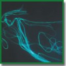
Results. Collagen autofluorescence has been registered in both fixed and non-fixed tissue graft samples, autofluorescence level of single fibers reflecting the extent of their compaction. Damage to the initial collagen structure leads to changes in tissue graft composition due to formation of picrinophilic fibers with very high autofluorescence levels, which do not appear in normal tissues or after collagen decondensation in conditions of hypotonia. Grafts with collagen fiber autofluorescence intensity 30 foot-candles are highly adhesive for M-22 human fibroblasts and donor platelets, whereas grafts with autofluorescence levels of more than 40 foot-candles exhibit significantly lower adhesive properties.
Conclusion. Collagen autofluorescence analysis provides important data about connective tissue topography, intercellular matrix integrity and allows collagen defects to be revealed, even in non-fixed tissue slices and grafts.
- Yermolov A., Macedonian T., Radygina M., Yartsev P., Titov G., Petukhov M., Papanino A., Kislitsina O., Andreyev Yu. Evaluation of the possibility of using tissue grafts in abdominal surgery. Khirurg 2015; 2: 47–60.
- Karmadonov A.V., Podolughny V.I., Zaicov I.N. Using modified xenopericardium in “non-strained plasties” of inferior abdominal wall hernia. Sibirskiy meditsinskiy zhurnal (Tomsk) 2008; 23(2): 28–32.
- Aleksandrov V.N., Khubulava G.G., Levanovich V.V. Tissue-engineered vascular grafts. Pediatr 2015; 6(1): 87–95.
- Khvatov V.B., Svishchev A.V., Vaza A.Yu., Borovkova N.V., Mironov A.S., Pokhitonov D.Yu., Andreev Yu.V. Method of manufacturing a lyophilized allograft bone. Transplantologiya 2016; 1: 13–18.
- Schug-Pass C., Sommerer F., Tannapfel A., Lippert H., Köckerling F. The use of composite meshes in laparoscopic repair of abdominal wall hernias: are there differences in biocompatibily? Surg Endosc 2008; 23(3): 487–495, https://doi.org/10.1007/s00464-008-0085-8.
- Bobrova M.M., Safonova L.А., Agapova О.I., Krasheninnikov M.E., Shagidulin M.Yu., Agapov I.I. Liver tissue decellularization as a promising porous scaffold processing technology for tissue engineering and regenerative medicine. Sovremennye tehnologii v medicine 2015; 7(4): 6–13, https://doi.org/10.17691/stm2015.7.4.01.
- Pokhitonov D.Y., Borovkova N.V., Filippov O.P., Klyukvin I.Y., Khvatov V.B., Ponomaryov I.N., Shugai S.V., Andreev Y.V., Smirnov S.V., Zhirkova E.A. Experimental substantiation and clinical use of a combination of dermal matrix with allogenic or autologous cells for the treatment of extensive traumatic wounds. Bull Exp Biol Med 2014; 157(5): 705–710, https://doi.org/10.1007/s10517-014-2647-1.
- Monteiro G.A., Rodriguez N.L., Delossantos A.I., Wagner C.T. Short-term in vivo biological and mechanical remodeling of porcine acellular dermal matrices. J Tissue Eng 2013; 4: 204173141349018, https://doi.org/10.1177/2041731413490182.
- Lamme E.N., de Vries H.J., van Veen H., Gabbiani G., Westerhof W., Middelkoop E. Extracellular matrix characterization during healing of full-thickness wounds treated with a collagen/elastin dermal substitute shows improved skin regeneration in pigs. J Histochem Cytochem 1996; 44(11): 1311–1322, https://doi.org/10.1177/44.11.8918906.
- Ahlfors J.-E.W., Billiar K.L. Biomechanical and biochemical characteristics of a human fibroblast-produced and remodeled matrix. Biomaterials 2007; 28(13): 2183–2191, https://doi.org/10.1016/j.biomaterials.2006.12.030.
- Jiang H., Rhee S., Ho C.-H., Grinnell F. Distinguishing fibroblast promigratory and procontractile growth factor environments in 3-D collagen matrices. FASEB J 2008; 22(7): 2151–2160, https://doi.org/10.1096/fj.07-097014.
- Pedersen J.A., Boschetti F., Swartz M.A. Effects of extracellular fiber architecture on cell membrane shear stress in a 3D fibrous matrix. J Biomech 2007; 40(7): 1484–1492, https://doi.org/10.1016/j.jbiomech.2006.06.023.
- Richards-Kortum R., Sevick-Muraca E. Quantitative optical spectroscopy for tissue diagnosis. Annu Rev Phys Chem 1996; 47(1): 555–606, https://doi.org/10.1146/annurev.physchem.47.1.555.
- Monici M. Cell and tissue autofluorescence research and diagnostic applications. Biotechnol Annu Rev 2005; 227–256, https://doi.org/10.1016/s1387-2656(05)11007-2.
- Baschong W., Suetterlin R., Laeng R.H. Control of autofluorescence of archival formaldehyde-fixed, paraffin-embedded tissue in confocal laser scanning microscopy (CLSM). J Histochem Cytochem 2001; 49(12): 1565–1571, https://doi.org/10.1177/002215540104901210.
- Khubutija M.Sh., Andreev J.V., Borovkova N.V., Khvatov V.B., Mironov A.S., Zhirkova E.A., Ponomarev I.N., Volkov K.S., Shugaj S.V., Konjushko O.I., Makarov M.S. Sposob izgotovleniya dermal’nogo matriksa [Method for making dermal matrix]. Patent RU 2524619. 2014.
- Makarov M.S., Khvatov V.B., Konjushko O.I., Borovkova N.V., Storozheva M.V., Ponomarev I.N. Metod morfofunktsional’noy otsenki kletochnogo komponenta biotransplantatov [Method for morphofunctional assessment of cell component of biografts]. Patent RU 2484472. 2013.
- Makarov M.S., Kobzeva E.N., Vysochin I.V., Borovkova N.V., Khvatov V.T. Morphofunctional analysis of human platelets by vital staining. Bull Exp Biol Med 2014; 156(3): 409–412, https://doi.org/10.1007/s10517-014-2360-0.
- Roskin G.I., Levinson L.B. Mikroskopicheskaya tekhnika [Microscopic technique]. Moscow: Sovetskaya nauka, 1957; 470 p.
- Makarov M.S., Storozheva M.V., Konyushko O.I., Borovkova N.V., Khvatov V.B. Effect of concentration of platelet-derived growth factor on proliferative activity of human fibroblasts. Bull Exp Biol Med 2013; 155(4): 576–580, https://doi.org/10.1007/s10517-013-2199-9.


