The Role of Functional MRI in Understanding the Origin of Speech Delay in Autism Spectrum Disorders
Autism spectrum disorders (ASD) are disorders of psychic development characterized by the difficulties of social interaction and stereotyped and repetitive patterns of behavior. Rather often they are accompanied by disturbances of speech, intelligence, and adaptive behavior. Pathogenesis of ASD is still poorly studied. MRI with its latest modalities is a modern diagnostic method enabling medical providers to evaluate structural, metabolic, and functional features of brain development in this pathology.
The aim of the study was to assess the capabilities of functional MRI (fMRI) in determining pathophysiological mechanisms of delay in speech development in ASD.
Materials and Methods. A brief review of international studies is given in the article. Our own results of examining 6 preschool children with one of the ASD forms — early childhood autism and speech disorders, and 6 children of the comparison group without autism and language disturbances are also presented using fMRI and a block design paradigm to analyze speech perception patterns.
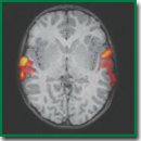
Results. In all children with normal speech development, bilateral symmetric spread of activation along the cortex of the entire superior temporal gyri was revealed whereas children with autism showed lateralized and limited involvement of the auditory cortex. Sevoflurane anesthesia did not influence the character of auditory zone activation.
Conclusion. The possibility of using fMRI with application of the paradigm for speech understanding to study the individual features of brain functioning in children with autism has been demonstrated. The revealed objective instrumental signs of brain activity differences in the children with autism compared to the healthy children allow the fMRI data to be considered as a potential biomarker of this disease. It has also been shown that the possibility to carry out this examination under general anesthesia makes it more acceptable and convenient for patients with childhood autism.
- Simashkova N.V., Makushkin E.V. Rasstroystva autisticheskogo spektra: diagnostika, lechenie, nablyudenie. Klinicheskie rekomendatsii (protokol lecheniya) [Autism spectrum disorders: diagnosis, treatment, monitoring. Clinical guidelines (treatment protocol)]. 2015.
- American Psychiatric Association. Diagnostic and statistical manual of mental disorders. American Psychiatric Association; 2013, https://doi.org/10.1176/appi.books.9780890425596.
- Ecker C., Spooren W., Murphy D.G.M. Translational approaches to the biology of autism: false dawn or a new era? Mol Psychiatry 2012; 18(4): 435–442, https://doi.org/10.1038/mp.2012.102.
- Haigh S.M., Heeger D.J., Dinstein I., Minshew N., Behrmann M. Cortical variability in the sensory-evoked response in autism. J Autism Dev Disord 2014; 45(5): 1176–1190, https://doi.org/10.1007/s10803-014-2276-6.
- Geschwind D.H., Levitt P. Autism spectrum disorders: developmental disconnection syndromes. Curr Opin Neurobiol 2007; 17(1): 103–111, https://doi.org/10.1016/j.conb.2007.01.009.
- Ha S., Sohn I.-J., Kim N., Sim H.J., Cheon K.-A. Characteristics of brains in autism spectrum disorder: structure, function and connectivity across the lifespan. Exp Neurobiol 2015; 24(4): 273, https://doi.org/10.5607/en.2015.24.4.273.
- Bernhardt B.C., Di Martino A., Valk S.L., Wallace G.L. Neuroimaging-based phenotyping of the autism spectrum. Curr Top Behav Neurosci 2017; 30: 341–355, https://doi.org/10.1007/7854_2016_438.
- Berg A.T., Dobyns W.B. Progress in autism and related disorders of brain development. Lancet Neurol 2015; 14(11): 1069–1070, https://doi.org/10.1016/s1474-4422(15)00048-4.
- Ismail M.M., Keynton R.S., Mostapha M.M., ElTanboly A.H., Casanova M.F., Gimel’farb G.L., El-Baz A. Studying autism spectrum disorder with structural and diffusion magnetic resonance imaging: a survey. Front Hum Neurosci 2016; 10: 211, https://doi.org/10.3389/fnhum.2016.00211.
- Heeger D.J., Ress D. What does fMRI tell us about neuronal activity? Nat Rev Neurosci 2002; 3(2): 142–151, https://doi.org/10.1038/nrn730.
- Logothetis N.K., Wandell B.A. Interpreting the BOLD signal. Annu Rev Physiol 2004; 66(1): 735–769, https://doi.org/10.1146/annurev.physiol.66.082602.092845.
- Logothetis N.K. The neural basis of the blood–oxygen–level–dependent functional magnetic resonance imaging signal. Philos Trans R Soc Lond B Biol Sci 2002; 357(1424): 1003–1037, https://doi.org/10.1098/rstb.2002.1114.
- Seliverstova E.V., Seliverstov Yu.A., Konovalov R.N., Illarioshkin S.N. Resting-state fMRI: new possibilities for studying physiology and pathology of the brain. Annaly kliniceskoj i eksperimental’noj nevrologii 2013; 7(4): 39–44.
- Seliverstov Y.A., Seliverstova E.V., Konovalov R.N., Klyushnikov S.A., Krotenkova M.V., Illarioshkin S.N. Clinical and imaging analysis of huntington disease with use of resting-state functional magnetic resonance imaging. The Neurological Journal 2015; 20(3): 11, https://doi.org/10.18821/1560-9545-2015-20-3-11-21.
- Dichter G.S. Functional magnetic resonance imaging of autism spectrum disorders. Dialogues Clin Neurosci 2012; 14(3): 319–351.
- Failla M.D., Moana-Filho E.J., Essick G.K., Baranek G.T., Rogers B.P., Cascio C.J. Initially intact neural responses to pain in autism are diminished during sustained pain. Autism 2017; 22(6): 669–683, https://doi.org/10.1177/1362361317696043.
- Gu X., Zhou T.J., Anagnostou E., Soorya L., Kolevzon A., Hof P.R., Fan J. Heightened brain response to pain anticipation in high-functioning adults with autism spectrum disorder. Eur J Neurosci 2017; 47(6): 592–601, https://doi.org/10.1111/ejn.13598.
- Carlisi C.O., Norman L., Murphy C.M., Christakou A., Chantiluke K., Giampietro V., Simmons A., Brammer M., Murphy D.G., Mataix-Cols D., Rubia K.; MRC AIMS consortium. Shared and disorder-specific neurocomputational mechanisms of decision-making in autism spectrum disorder and obsessive-compulsive disorder. Cereb Cortex 2017; 27(12): 5804–5816, https://doi.org/10.1093/cercor/bhx265.
- Stanfield A.C., Philip R.C.M., Whalley H., Romaniuk L., Hall J., Johnstone E.C., Lawrie S.M. Dissociation of brain activation in autism and schizotypal personality disorder during social judgments. Schizophr Bull 2017; 43(6): 1220–1228, https://doi.org/10.1093/schbul/sbx083.
- Murphy C.M., Christakou A., Giampietro V., Brammer M., Daly E.M., Ecker C., Johnston P., Spain D., Robertson D.M.; MRC AIMS Consortium, Murphy D.G., Rubia K. Abnormal functional activation and maturation of ventromedial prefrontal cortex and cerebellum during temporal discounting in autism spectrum disorder. Hum Brain Mapp 2017; 38(11): 5343–5355,https://doi.org/10.1002/hbm.23718.
- Philip R.C., Dauvermann M.R., Whalley H.C., Baynham K., Lawrie S.M., Stanfield A.C. A systematic review and meta-analysis of the fMRI investigation of autism spectrum disorders. Neurosci Biobehav Rev 2012; 36(2): 901–942, https://doi.org/10.1016/j.neubiorev.2011.10.008.
- Kim S.Y., Choi U.S., Park S.Y., Oh S.H., Yoon H.W., Koh Y.J., Im W.Y., Park J.I., Song D.H., Cheon K.A., Lee C.U. Abnormal activation of the social brain network in children with autism spectrum disorder: an fMRI study. Psychiatry Investig 2015; 12(1): 37, https://doi.org/10.4306/pi.2015.12.1.37.
- Di Martino A., Ross K., Uddin L.Q., Sklar A.B., Castellanos F.X., Milham M.P. Functional brain correlates of social and nonsocial processes in autism spectrum disorders: an activation likelihood estimation meta-analysis. Biol Psychiatry 2009; 65(1): 63–74, https://doi.org/10.1016/j.biopsych.2008.09.022.
- Lombardo M.V., Pierce K., Eyler L.T., Carter Barnes C., Ahrens-Barbeau C., Solso S., Campbell K., Courchesne E. Different functional neural substrates for good and poor language outcome in autism. Neuron 2015; 86(2): 567–577, https://doi.org/10.1016/j.neuron.2015.03.023.
- Wang A.T., Lee S.S., Sigman M., Dapretto M. Neural basis of irony comprehension in children with autism: the role of prosody and context. Brain 2006; 129(4): 932–943, https://doi.org/10.1093/brain/awl032.
- Redcay E., Courchesne E. Deviant functional magnetic resonance imaging patterns of brain activity to speech in 2–3-year-old children with autism spectrum disorder. Biol Psychiatry 2008; 64(7): 589–598, https://doi.org/10.1016/j.biopsych.2008.05.020.
- Kana R.K., Keller T.A., Cherkassky V.L., Minshew N.J., Just M.A. Sentence comprehension in autism: thinking in pictures with decreased functional connectivity. Brain 2006; 129(9): 2484–2493, https://doi.org/10.1093/brain/awl164.
- Lee Y., Park B., James O., Kim S.-G., Park H. Autism spectrum disorder related functional connectivity changes in the language network in children, adolescents and adults. Front Hum Neurosci 2017; 11: 418, https://doi.org/10.3389/fnhum.2017.00418.
- Chouinard B., Volden J., Cribben I., Cummine J. Neurological evaluation of the selection stage of metaphor comprehension in individuals with and without autism spectrum disorder. Neuroscience 2017; 361: 19–33, https://doi.org/10.1016/j.neuroscience.2017.08.001.
- Knaus T.A., Burns C., Kamps J., Foundas A.L. Atypical activation of action-semantic network in adolescents with autism spectrum disorder. Brain Cogn 2017; 117: 57–64, https://doi.org/10.1016/j.bandc.2017.06.004.
- Yang J., Lee J. Different aberrant mentalizing networks in males and females with autism spectrum disorders: evidence from resting-state functional magnetic resonance imaging. Autism 2016; 22(2): 134–148, https://doi.org/10.1177/1362361316667056.
- Lenroot R.K., Yeung P.K. Heterogeneity within autism spectrum disorders: what have we learned from neuroimaging studies? Front Hum Neurosci 2013; 7, https://doi.org/10.3389/fnhum.2013.00733.
- Andrews D.S., Marquand A., Ecker C., McAlonan G. Using pattern classification to identify brain imaging markers in autism spectrum disorder. Curr Top Behav Neurosci 2018; 40: 413–436, https://doi.org/10.1007/7854_2018_47.
- Redcay E. The superior temporal sulcus performs a common function for social and speech perception: implications for the emergence of autism. Neurosci Biobehav Rev 2008; 32(1): 123–142, https://doi.org/10.1016/j.neubiorev.2007.06.004.
- Pierce K. Early functional brain development in autism and the promise of sleep fMRI. Brain Res 2011; 1380: 162–174, https://doi.org/10.1016/j.brainres.2010.09.028.
- Tang C.Y., Ramani R. fMRI and anesthesia. Int Anesthesiol Clin 2016; 54(1): 129–142, https://doi.org/10.1097/aia.0000000000000081.
- Gemma M., Scola E., Baldoli C., Mucchetti M., Pontesilli S., De Vitis A., Falini A., Beretta L. Auditory functional magnetic resonance in awake (nonsedated) and propofol-sedated children. Paediatr Anaesth 2016; 26(5): 521–530, https://doi.org/10.1111/pan.12884.
- Gemma M., Scola E., Baldoli C., Mucchetti M., Pontesilli S., De Vitis A., Falini A., Beretta L. Brain resting-state functional connectivity is preserved under sevoflurane anesthesia in patients with pervasive developmental disorders: a pilot study. Brain Connect 2017; 7(4): 250–257, https://doi.org/10.1089/brain.2016.0448.
- Raschle N.M., Smith S.A., Zuk J., Dauvermann M.R., Figuccio M.J., Gaab N. Investigating the neural correlates of voice versus speech-sound directed information in pre-school children. PLoS One 2014; 9(12): e115549, https://doi.org/10.1371/journal.pone.0115549.
- Markou P., Ahtam B., Papadatou-Pastou M. Elevated levels of atypical handedness in autism: meta-analyses. Neuropsychol Rev 2017; 27(3): 258–283, https://doi.org/10.1007/s11065-017-9354-4.
- Paquet A., Golse B., Girard M., Olliac B., Vaivre-Douret L. Laterality and lateralization in autism spectrum disorder, using a standardized neuro-psychomotor assessment. Dev Neuropsychol 2017; 42(1): 39–54, https://doi.org/10.1080/87565641.2016.1274317.


