Indications for the Use of Sialoendoscopy in Sialolithiasis
The aim of the study is to determine indications for the use of sialoendoscopy in the diagnosis and treatment of sialolithiasis.
Materials and Methods. The study involved 115 patients with sialolithiasis, who underwent cone beam computed tomography, ultrasound diagnosis of the salivary glands, and sialoendoscopy, in addition to the standard general clinical examination.
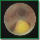
Results. Sialoendoscopy makes it possible to detect a stone, determine its shape, relative size, mobility, and assess the condition of the salivary ducts. It is impossible to obtain this information by other methods, though it is very important for treatment decision making. The design of the sialoscope and its special instruments make it possible to proceed with sialolith extraction immediately after detecting it.
Conclusion. The absolute indication for the use of sialoendoscopy is mobile calculi less than 5 mm in diameter (L1 according to F. Marchal’s LSD classification). In case of immobile sialoliths less than 4–8 mm in size, located in the main duct (L2), endoscopy should be used as a method supplementary to ductotomy. When sialoliths are located in the distal parts behind the areas of bending or stricture (L3a and L3b), the use of endoscopy is not indicated.
- Chechina I.N. Otsenka effektivnosti konservativnogo lecheniya sialolitiaza. Avtoref. dis. … kand. med. nauk [Evaluation of the effectiveness of conservative treatment of sialolithiasis. PhD Thesis]. Barnaul; 2010.
- Koch M., Zenk J., Iro H. Speichelgangsendoskopie in der Diagnostik und Therapie von obstruktiven Speicheldrüsenerkrankungen. HNO 2008; 56(2): 139–144, https://doi.org/10.1007/s00106-007-1563-3.
- Marchal F., Dulguerov P. Sialolithiasis management: the state of the art. Arch Otolaryngol Head Neck Surg 2003; 129(9): 951–916, https://doi.org/10.1001/archotol.129.9.951.
- Nahlieli O. Modern management preserving the salivary glands. J Oral Maxillofac Surg 2009; 67(9): 114–115, https://doi.org/10.1016/j.joms.2009.05.212.
- Katz P. New method of examination of the salivary glands: the fiberscope. Inf Dent 1990; 72(10): 785–786.
- Katz P. New therapy for sialolithiasis. Inf Dent 1991; 73(43): 3975–3979.
- Katz P. Endoscopy of the salivary glands. Ann Radiol (Paris) 1991; 34(1–2): 110–113.
- Rzymska-Grala I., Stopa Z., Grala B., Gołębiowski M., Wanyura H., Zuchowska A., Sawicka M., Zmorzyński M. Salivary gland calculi — contemporary methods of imaging. Pol J Radiol 2010; 75(3): 25–37.
- Vaiman M. Comparative analysis of methods of endoscopic surgery of the submandibular gland: 114 surgeries. Clin Otolaryngol 2015; 40(2): 162–166, https://doi.org/10.1111/coa.12357.
- Marchal F., Dulguerov P., Becker M., Barki G., Disant F., Lehmann W. Submandibular diagnostic and interventional sialendoscopy: new procedure for ductal disorders. Ann Otol Rhinol Laryngol 2002; 111(1): 27–35, https://doi.org/10.1177/000348940211100105.
- Marchal F. Endoscopie des canaux salivaires: toujours plus petit, toujours plus loin? Rev Stomatol Chir Maxillofac 2005; 106(4): 244–249, https://doi.org/10.1016/s0035-1768(05)85853-x.
- Marchal F., Kurt M., Dulguerov P., Becker M., Oedman M., Lehmann W. Histopathology of submandibular glands removed for sialolithiasis. Ann Otol Rhinol Laryngol 2001; 110(5 Pt 1): 464–469, https://doi.org/10.1177/000348940111000513.
- Nahlieli O., Baruchin A.M. Endoscopic technique for the diagnosis and treatment of obstructive salivary gland diseases. J Oral Maxillofac Surg 1999; 57(12): 1394–1402, https://doi.org/10.1016/s0278-2391(99)90716-4.
- Strychowsky J.E., Sommer D.D., Gupta M.K., Cohen N., Nahlieli O. Sialendoscopy for the management of obstructive salivary gland disease: a systematic review and meta-analysis. Arch Otolaryngol Head Neck Surg 2012; 138(6): 541–547, https://doi.org/10.1001/archoto.2012.856.
- Koch M., Zenk J., Bozzatto A., Bumm K., Iro H. Sialoscopy in cases of unclear swelling of the major salivary glands. Otolaryngol Head Neck Surg 2005; 133(6): 863–868, https://doi.org/10.1016/j.otohns.2005.08.005.


