Мультимодальная оптическая когерентная томография как метод визуализации состояния нервной ткани при глиальных опухолях головного мозга (экспериментальное исследование)
Цель исследования — изучение возможностей мультимодальной ОКТ (ММ ОКТ) в дифференциации нормальной и патологически измененной ткани головного мозга на примере экспериментальной модели глиобластомы.
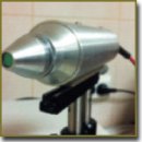
Материалы и методы. Исследования выполнены на экспериментальной установке скоростной спектральной ММ ОКТ, разработанной в ИПФ РАН (Н. Новгород) и включающей два режима исследования: кросс-поляризационную ОКТ (КП ОКТ) и спектральную микроангиографическую ОКТ (МА ОКТ). Характеристики установки: скорость получения информации — 20 000 А-сканов в секунду; длина волны — 1,3 мкм; размер кадра ~4×2 мм; поперечное разрешение — 20 мкм; разрешение по глубине — 10–15 мкм. ОКТ-исследование проведено на экспериментальной опухолевой модели глиобластомы крысы 101.8, которая была привита и поддерживается в Институте морфологии человека. С целью оценки параметров сигнала, характерных для опухоли и неизмененной мозговой ткани, КП ОКТ- и МА ОКТ-изображения сопоставляли с гистологическими препаратами (окраска гематоксилином и эозином). Анализ МА ОКТ-изображений также проводили на основании сравнения с данными ZOOM-микроскопии.
Результаты. На примере модели глиобластомы крысы 101.8 установлена связь характера КП ОКТ-изображений участков ткани головного мозга с их морфологической структурой. Произведена сравнительная оценка сигналов от глиальной опухоли и неизмененной мозговой ткани. МА ОКТ позволяет визуализировать опухолевые и нормальные мозговые сосуды, выявляя характерные изменения формы и размеров сосудов опухоли.
Заключение. ММ ОКТ является инновационной технологией, перспективной для использования в качестве метода интраоперационной диагностики при глиальных опухолях головного мозга. Возможность сочетать несколько режимов исследования позволяет одновременно получать информацию как о структуре ткани, так и об особенностях строения микрососудистой сети и ее элементов.
- Witiw C.D., Nathan V., Bernstein M. Economics, innovation, and quality improvement in neurosurgery. Neurosurg Clin N Am 2015; 26(2): 197–205, http://dx.doi.org/10.1016/j.nec.2014.11.003.
- Moiseev A.A., Gelikonov G.V., Terpelov D.A., Shilyagin P.A., Gelikonov V.M. Improvement of lateral resolution of spectral domain optical coherence tomography images in out-of-focus regions with holographic data processing techniques. Quantum Electronics 2014; 44(8): 732–739, http://dx.doi.org/10.1070/QE2014v044n08ABEH015492.
- Gelikonov V.M., Gelikonov G.V., Shilyagin P.A. Linear-wavenumber spectrometer for high-speed spectral-domain optical coherence tomography. Optics and Spectroscopy 2009; 106(3): 459–465, http://dx.doi.org/10.1134/s0030400x09030242.
- Gelikonov V.M., Gelikonov G.V., Kasatkina I.V., Terpelov D.A., Shilyagin P.A. Coherent noise compensation in spectral-domain optical coherence tomography. Optics and Spectroscopy 2009; 106(6): 895–900, http://dx.doi.org/10.1134/s0030400x09060174.
- Gelikonov V.M., Gelikonov G.V., Terpelov D.A., Shabanov D.V., Shilyagin P.A. Suppression of image autocorrelation artefacts in spectral domain optical coherence tomography and multiwave digital holography. Quantum Electronics 2012; 42(5): 390–393, http://dx.doi.org/10.1070/QE2012v042n05ABEH014852.
- Shilyagin P.A., Gelikonov G.V., Gelikonov V.M., Moiseev A.A., Terpelov D.A. Achromatic registration of quadrature components of the optical spectrum in spectral domain optical coherence tomography. Quantum Electronics 2014; 44(7): 664–669, http://dx.doi.org/10.1070/QE2014v044n07ABEH015465.
- Gelikonov V.M., Gelikonov G.V. New approach to cross-polarized optical coherence tomography based on orthogonal arbitrarily polarized modes. Laser Physics Letters 2006; 3(9): 445–451, http://dx.doi.org/10.1002/lapl.200610030.
- Matveev L.A., Zaitsev V.Y., Gelikonov G.V., Matveyev A.L., Moiseev A.A., Ksenofontov S.Y., Gelikonov V.M., Sirotkina M.A., Gladkova N.D., Demidov V., Vitkin A. Hybrid M-mode-like OCT imaging of three-dimensional microvasculature in vivo using reference-free processing of complex valued B-scans. Optics Letters 2015; 40(7): 1472–1475, http://dx.doi.org/10.1364/ol.40.001472.
- Халанский А.С., Кондакова Л.И. Перевиваемый штамм глиомы крысы 101.8.I. Биологическая характеристика. Клиническая и экспериментальная морфология 2013; 4(8): 63–68.
- Barth R.F., Kaur B. Rat brain tumor models in experimental neuro-oncology: the C6, 9L, T9, RG2, F98, BT4C, RT-2 and CNS-1 gliomas. J Neurooncol 2009; 94(3): 299–312, http://dx.doi.org/10.1007/s11060-009-9875-7.
- Goldey G.J., Roumis D.K., Glickfeld L.L., Kerlin A.M., Reid R.C., Bonin V., Schafer D.P., Andermann M.L. Removable cranial windows for long-term imaging in awake mice. Nat Protoc 2014; 9(11): 2515–2538, http://dx.doi.org/10.1038/nprot.2014.165.
- Mostany R., Portera-Cailliau C. A craniotomy surgery procedure for chronic brain imaging. J Vis Exp 2008; 12, http://dx.doi.org/10.3791/680.
- Park K., You J., Du C., Pan Y. Cranial window implantation on mouse cortex to study microvascular change induced by cocaine. Quant Imaging Med Surg 2015; 5(1): 97–107, http://dx.doi.org/10.3978/j.issn.2223-4292.2014.11.31.
- Keiner D., Heimann A., Kronfeld A., Sommer C., Mueller-Forell W., Kempski O., Oertel J. Towards a glioma model for surgical technique evaluation in the rat. Br J Neurosurg 2014; 28(1): 86–92, http://dx.doi.org/10.3109/02688697.2013.804489.
- Приказ Минздравсоцразвития РФ от 23.08.2010 №708н “Об утверждении Правил лабораторной практики”.
- Международные рекомендации (этический кодекс) по проведению медико-биологических исследований с использованием животных. 1985.
- Sanai N., Berger M.S. Glioma extent of resection and its impact on patient outcome. Neurosurgery 2008; 62(4): 753–766, http://dx.doi.org/10.1227/01.neu.0000318159.21731.cf.
- Sanai N., Polley M.Y., McDermott M.W., Parsa A.T., Berger M.S. An extent of resection threshold for newly diagnosed glioblastomas. J Neurosurg 2011; 115(1): 3–8, http://dx.doi.org/10.3171/2011.2.jns10998.
- Анохина Ю.Е. Гайдар Б.В., Мартынов Б.В., Свистов Д.В., Папаян Г.В., Григорьевский Д.И. Прогностическая значимость объема хирургического вмешательства в условиях применения интраоперационной флуоресцентной диагностики у пациентов со злокачественными глиомами головного мозга. Вестник российской военно-медицинской академии 2014; 1: 19–24.
- Stummer W., Reulen H.J., Meinel T., Pichlmeier U., Schumacher W., Tonn J.C., Rohde V., Oppel F., Turowski B., Woiciechowsky C., Franz K., Pietsch T. Extent of resection and survival in glioblastoma multiforme: identification of and adjustment for bias. Neurosurgery 2008; 62(3): 564–576, http://dx.doi.org/10.1227/01.neu.0000317304.31579.17.
- McGirt M.J., Chaichana K.L., Gathinji M., Attenello F.J., Than K., Olivi A., Weingart J.D., Brem H., Quiñones-Hinojosa A.R. Independent association of extent of resection with survival in patients with malignant brain astrocytoma. J Neurosurg 2009; 110(1): 156–162, http://dx.doi.org/10.3171/2008.4.17536.
- Kuhnt D., Becker A., Ganslandt O., Bauer M., Buchfelder M., Nimsky C. Correlation of the extent of tumor volume resection and patient survival in surgery of glioblastoma multiforme with high-field intraoperative MRI guidance. Neuro Oncol 2011; 13(12): 1339–1348, http://dx.doi.org/10.1093/neuonc/nor133.
- Sanai N., Polley M.Y., McDermott M.W., Parsa A.T., Berger M.S. An extent of resection threshold for newly diagnosed glioblastomas. J Neurosurg 2011; 115(1): 3–8, http://dx.doi.org/10.3171/2011.2.JNS10998.
- Böhringer H.J., Boller D., Leppert J., Knopp U., Lankenau E., Reusche E., Hüttmann G., Giese A. Time-domain and spectral-domain optical coherence tomography in the analysis of brain tumor tissue. Lasers Surg Med 2006; 38(6): 588–597, http://dx.doi.org/10.1002/lsm.20353.
- Böhringer H.J., Lankenau E., Stellmacher F., Reusche E., Hüttmann G., Giese A. Imaging of human brain tumor tissue by near-infrared laser coherence tomography. Acta Neurochir (Wien) 2009; 151(5): 507–517, http://dx.doi.org/10.1007/s00701-009-0248-y.
- Kut C., Chaichana K.L., Xi J., Raza S.M., Ye X., McVeigh E.R., Rodriguez F.J., Quiñones-Hinojosa A., Li X. Detection of human brain cancer infiltration ex vivo and in vivo using quantitative optical coherence tomography. Sci Transl Med 2015; 7(292): 292ra100, http://dx.doi.org/10.1126/scitranslmed.3010611.
- Kantelhardt S.R., Finke M., Schweikard A., Giese A. Evaluation of a completely robotized neurosurgical operating microscope. Neurosurgery 2013; 72(Suppl 1): A19–A26, http://dx.doi.org/10.1227/NEU.0b013e31827235f8.
- Finke M., Kantelhardt S., Schlaefer A., Bruder R., Lankenau E., Giese A., Schweikard A. Automatic scanning of large tissue areas in neurosurgery using optical coherence tomography. Int J Med Robot 2012; 8(3): 327–336, http://dx.doi.org/10.1002/rcs.1425.
- Lankenau E., Klinger D., Winter C., Malik A., Müller H., Oelckers S., Pau H.-W., Just T., Hüttmann G. Combining optical coherence tomography (OCT) with an operating microscope. In: Advances in medical engineering. Springer Berlin Heidelberg; 2007, p. 343–348, http://dx.doi.org/10.1007/978-3-540-68764-1_57.
- Schmitt J.M., Xiang S.H. Cross-polarized backscatter in optical coherence tomography of biological tissue. Opt Lett 1998; 23(13): 1060–1062, http://dx.doi.org/10.1364/ol.23.001060.
- Gladkova N., Kiseleva E., Streltsova O., Prodanets N., Snopova L., Karabut M., Gubarkova E., Zagaynova E. Combined use of fluorescence cystoscopy and cross-polarization OCT for diagnosis of bladder cancer and correlation with immunohistochemical markers. J Biophotonics 2013; 6(9): 687–698, http://dx.doi.org/10.1002/jbio.201200105.
- Gladkova N., Streltsova O., Zagaynova E., Kiseleva E., Gelikonov V., Gelikonov G., Karabut M., Yunusova K., Evdokimova O. Cross-polarization optical coherence tomography for early bladder-cancer detection: statistical study. J Biophotonics 2011; 4(7–8): 519–532, http://dx.doi.org/10.1002/jbio.201000088.
- Gladkova N., Kiseleva E., Robakidze N., Balalaeva I., Karabut M., Gubarkova E., Feldchtein F. Evaluation of oral mucosa collagen condition with cross-polarization optical coherence tomography. J Biophotonics 2013; 6(4): 321–329, http://dx.doi.org/10.1002/jbio.201200059.
- Kiseleva E., Kirillin M., Feldchtein F., Vitkin A., Sergeeva E., Zagaynova E., Streltzova O., Shakhov B., Gubarkova E., Gladkova N. Differential diagnosis of human bladder mucosa pathologies in vivo with cross-polarization optical coherence tomography. Biomed Opt Express 2015; 6(4):1464–1476, http://dx.doi.org/10.1364/boe.6.001464.
- Leitgeb R.A., Werkmeister R.M., Blatter C., Schmetterer L. Doppler optical coherence tomography. Prog Retin Eye Res 2014; 41: 26–43, http://dx.doi.org/10.1016/j.preteyeres.2014.03.004.
- Chong S.P., Merkle C.W., Leahy C., Srinivasan V.J. Cerebral metabolic rate of oxygen (CMRO2) assessed by combined Doppler and spectroscopic OCT. Biomed Opt Express 2015; 6(10): 3941–3951, http://dx.doi.org/10.1364/BOE.6.003941.
- Devor A., Sakadzic S., Srinivasan V.J., Yaseen M.A., Nizar K., Saisan P.A., Tian P., Dale A.M., Vinogradov S.A., Franceschini M.A., Boas D.A. Frontiers in optical imaging of cerebral blood flow and metabolism. J Cereb Blood Flow Metab 2012; 32(7): 1259–1276, http://dx.doi.org/10.1038/jcbfm.2011.195.
- Srinivasan V.J., Sakadžić S., Gorczynska I., Ruvinskaya S., Wu W., Fujimoto J.G., Boas D.A. Quantitative cerebral blood flow with optical coherence tomography. Opt Express 2010; 18(3): 2477–2494, http://dx.doi.org/10.1364/OE.18.002477.
- Lee J., Wu W., Lesage F., Boas D.A. Multiple-capillary measurement of RBC speed, flux, and density with optical coherence tomography. J Cereb Blood Flow Metab 2013; 33(11): 1707–1710, http://dx.doi.org/10.1038/jcbfm.2013.158.
- Srinivasan V.J., Atochin D.N., Radhakrishnan H., Jiang J.Y., Ruvinskaya S., Wu W., Barry S., Cable A.E., Ayata C., Huang P.L., Boas D.A. Optical coherence tomography for the quantitative study of cerebrovascular physiology. J Cereb Blood Flow Metab 2011; 31(6): 1339–1345, http://dx.doi.org/10.1038/jcbfm.2011.19.
- Ren H., Du C., Yuan Z., Park K., Volkow N.D., Pan Y. Cocaine-induced cortical microischemia in the rodent brain: clinical implications. Mol Psychiatry 2012; 17(10): 1017–1025, http://dx.doi.org/10.1038/mp.2011.160.


