Role of Familiarity in Recognizing Faces and Words: an EEG Study
The ability to recognize faces and words is crucial for social communication. A close relationship between the face recognition and the word recognition has been documented in numerous studies involving EEG, evoked potentials, functional MRI, as well as clinical data in patients with impaired face perception who suffered from some word recognition inability. Therefore, identifying common mechanisms underlying the recognition of verbal and non-verbal stimuli is relevant not only for normal physiology and cognitive neuroscience, but also for the clinical conditions of agnosia of various etiologies.
We hypothesized that EEG patterns related to the recognition of words and faces might be influenced by both the stimulus-type factor (word or face) and the factor of familiarity.
The aim of the study is to identify the EEG patterns responsible for the perception and recognition of a visual stimulus, regardless of its specificity, i.e. the common patterns for verbal (words) and non-verbal (faces) stimuli.
Materials and Methods. The EEG data were obtained from 26 volunteers who were presented with complex visual stimuli, i.e., photos of people with words superimposed on them, where familiar (known) and unfamiliar people and words were combined in equal parts. Firstly, the tested subjects were asked to classify the faces into familiar or unfamiliar (with the attention only to faces); secondly — to classify the words in the same way (the attention only to words).
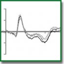
Results. We found a pronounced effect of familiarity on the EEG patterns: the amplitude of the N250 component of evoked potentials detected in the frontal areas was significantly greater in the responses to unfamiliar stimuli (both faces and words) compared to familiar ones. We also found an effect of instruction on the responses:the N400 component amplitude was greater in responses followed the “attention to words” instruction as compared to the “attention to faces” instruction; this effect was also best detectable in the frontal sites.
Conclusion. At the early stages of the visual stimuli recognition, the evoked potentials responses are modulated by the familiarity of the stimuli (that is, by their representation in long-term memory), and not by their type (face or word). The categorization of stimuli by their modality (verbal or non-verbal), apparently, occurs at later stages of their processing.
- Puce A., Allison T., Asgari M., Gore J.C., McCarthy G. Differential sensitivity of human visual cortex to faces, letter strings, and textures: a functional magnetic resonance imaging study. J Neurosci 1996; 16(16): 5205–5215, https://doi.org/10.1523/jneurosci.16-16-05205.1996.
- McCarthy G., Puce A., Gore J.C., Allison T. Face-specific processing in the human fusiform gyrus. J Cogn Neurosci 1997; 9(5): 605–610, https://doi.org/10.1162/jocn.1997.9.5.605.
- Cohen L., Dehaene S., Naccache L., Lehéricy S., Dehaene-Lambertz G., Hénaff M.A., Michel F. The visual word form area: spatial and temporal characterization of an initial stage of reading in normal subjects and posterior split-brain patients. Brain 2000; 123(2): 291–307, https://doi.org/10.1093/brain/123.2.291.
- Yovel G., Tambini A., Brandman T. The asymmetry of the fusiform face area is a stable individual characteristic that underlies the left-visual-field superiority for faces. Neuropsychologia 2008; 46(13): 3061–3068, https://doi.org/10.1016/j.neuropsychologia.2008.06.017.
- Dien J. A tale of two recognition systems: implications of the fusiform face area and the visual word form area for lateralized object recognition models. Neuropsychologia 2009; 47(1): 1–16, https://doi.org/10.1016/j.neuropsychologia.2008.08.024.
- Kanwisher N., McDermott J., Chun M.M. The fusiform face area: a module in human extrastriate cortex specialized for face perception. J Neurosci 1997; 17(11): 4302–4311, https://doi.org/10.1523/jneurosci.17-11-04302.1997.
- Bouhali F., de Schotten M.T., Pinel P., Poupon C., Mangin J.F., Dehaene S., Cohen L. Anatomical connections of the visual word form area. J Neurosci 2014; 34(46): 15402–15414, https://doi.org/10.1523/jneurosci.4918-13.2014.
- Harris R.J., Rice G.E., Young A.W., Andrews T.J. Distinct but overlapping patterns of response to words and faces in the fusiform gyrus. Cereb Cortex 2015; 26(7): 3161–3168, https://doi.org/10.1093/cercor/bhv147.
- Robinson A.K., Plaut D.C., Behrmann M. Word and face processing engage overlapping distributed networks: evidence from RSVP and EEG investigations. J Exp Psychol Gen 2017; 146(7): 943–961, https://doi.org/10.1037/xge0000302.
- Farah M.J. Cognitive neuropsychology: patterns of co-occurrence among the associative agnosias: implications for visual object representation. Cogn Neuropsychol 1991; 8(1): 1–19, https://doi.org/10.1080/02643299108253364.
- Farah M.J. Dissociable systems for visual recognition: a cognitive neuropsychology approach. In: Kosslyn S.M., Osherson D. (editors). Visual cognition: an invitation to cognitive science. Vol. 2. Cambridge: MIT Press; 1995; p. 101–119.
- Buxbaum L.J., Glosser G., Coslett H.B. Impaired face and word recognition without object agnosia. Neuropsychologia 1998; 37(1): 41–50, https://doi.org/10.1016/s0028-3932(98)00048-7.
- Geskin J., Behrmann M. Congenital prosopagnosia without object agnosia? A literature review. Cogn Neuropsychol 2018; 35(1–2): 4–54, https://doi.org/10.1080/02643294.2017.1392295.
- Behrmann M., Plaut D.C. Bilateral hemispheric processing of words and faces: evidence from word impairments in prosopagnosia and face impairments in pure alexia. Cereb Cortex 2012; 24(4): 1102–1118, https://doi.org/10.1093/cercor/bhs390.
- Rossion B., Jacques C. The N170: understanding the time course of face perception in the human brain. In: Luck S.J., Kappenman A.S. (editors). The Oxford handbook of event-related potential components. Oxford University Press; 2011; p. 15–141, https://doi.org/10.1093/oxfordhb/9780195374148.013.0064.
- Eimer M. Event-related brain potentials distinguish processing stages involved in face perception and recognition. Neurophysiol Clin 2000; 111(4): 694–705, https://doi.org/10.1016/s1388-2457(99)00285-0.
- Engst F.M., Martín-Loeches M., Sommer W. Memory systems for structural and semantic knowledge of faces and buildings. Brain Res 2006; 1124(1): 70–80, https://doi.org/10.1016/j.brainres.2006.09.038.
- Caharel S., Poiroux S., Bernard C., Thibaut F., Lalonde R., Rebai M. ERPs associated with familiarity and degree of familiarity during face recognition. Int J Neurosci 2002; 112(12): 1499–1512, https://doi.org/10.1080/00207450290158368.
- Caharel S., Fiori N., Bernard C., Lalonde R., Rebaï M. The effects of inversion and eye displacements of familiar and unknown faces on early and late-stage ERPs. Int J Psychophysiol 2006; 62(1): 141–151, https://doi.org/10.1016/j.ijpsycho.2006.03.002.
- Wild-Wall N., Dimigen O., Sommer W. Interaction of facial expressions and familiarity: ERP evidence. Biol Psychol 2008; 77(2): 138–149, https://doi.org/10.1016/j.biopsycho.2007.10.001.
- Bentin S., Mouchetant-Rostaing Y., Giard M.H., Echallier J.F., Pernier J. ERP manifestations of processing printed words at different psycholinguistic levels: time course and scalp distribution. J Cogn Neurosci 1999; 11(3): 235–260, https://doi.org/10.1162/089892999563373.
- Maurer U., Brandeis D., McCandliss B.D. Fast, visual specialization for reading in English revealed by the topography of the N170 ERP response. Behav Brain Funct 2005; 1(1): 13, https://doi.org/10.1186/1744-9081-1-13.
- Cao X., Jiang B., Gaspar C., Li C. The overlap of neural selectivity between faces and words: evidences from the N170 adaptation effect. Exp Brain Res 2014; 232(9): 3015–3021, https://doi.org/10.1007/s00221-014-3986-x.
- Zhao J., Li S., Lin S.E., Cao X.H., He S., Weng X.C. Selectivity of N170 in the left hemisphere as an electrophysiological marker for expertise in reading Chinese. Neurosci Bull 2012; 28(5): 577–584, https://doi.org/10.1007/s12264-012-1274-y.
- Rossion B., Joyce C.A., Cottrell G.W., Tarr M.J. Early lateralization and orientation tuning for face, word, and object processing in the visual cortex. Neuroimage 2003; 20(3): 1609–1624, https://doi.org/10.1016/j.neuroimage.2003.07.010.
- Leleu A., Caharel S., Carré J., Montalan B., Snoussi M., Vom Hofe A., Charvin H., Lalonde R., Rebaï M. Perceptual interactions between visual processing of facial familiarity and emotional expression: an event-related potentials study during task-switching. Neurosci Lett 2010; 482(2): 106–111, https://doi.org/10.1016/j.neulet.2010.07.008.
- Tanaka J.W., Curran T., Porterfield A.L., Collins D. Activation of preexisting and acquired face representations: the N250 event-related potential as an index of face familiarity. J Cogn Neurosci 2006; 18(9): 1488–1497, https://doi.org/10.1162/jocn.2006.18.9.1488.
- Begleiter H., Porjesz B., Wang W. Event-related brain potentials differentiate priming and recognition to familiar and unfamiliar faces. Electroencephalogr Clin Neurophysiol 1995; 94(1): 41–49, https://doi.org/10.1016/0013-4694(94)00240-L.
- Miyakoshi M., Nomura M., Ohira H. An ERP study on self-relevant object recognition. Brain Cogn 2007; 63(2): 182–189, https://doi.org/10.1016/j.bandc.2006.12.001.
- Itier R.J., Taylor M.J. Inversion and contrast polarity reversal affect both encoding and recognition processes of unfamiliar faces: a repetition study using ERPs. Neuroimage 2002; 15(2): 353–372, https://doi.org/10.1006/nimg.2001.0982.
- Bentin S., Deouell L.Y. Structural encoding and identification in face processing: ERP evidence for separate mechanisms. Cogn Neuropsychol 2000; 7(1–3): 35–55, https://doi.org/10.1080/026432900380472.
- Yick Y.Y., Wilding E.L. Material-specific neural correlates of memory retrieval. Neuroreport 2008; 19(15): 1463–1467, https://doi.org/10.1097/wnr.0b013e32830ef76f.
- MacKenzie G., Donaldson D.I. Examining the neural basis of episodic memory: ERP evidence that faces are recollected differently from names. Neuropsychologia 2009; 47(13): 2756–2765, https://doi.org/10.1016/j.neuropsychologia.2009.05.025.
- Joyce C.A., Kutas M. Event-related potential correlates of long-term memory for briefly presented faces. J Cogn Neurosci 2005; 17(5): 757–767, https://doi.org/10.1162/0898929053747603.
- Rugg M.D., Allan K. Event-related potential studies of memory. In: Tulving E., Craik F.I.M. (editors). The Oxford handbook of memory. Oxford University Press; 2000; p. 521–537.
- Meyer P., Mecklinger A., Friederici A.D. Bridging the gap between the semantic N400 and the early old/new memory effect. Neuroreport 2007; 18(10): 1009–1013, https://doi.org/10.1097/wnr.0b013e32815277eb.
- Kutas M., Federmeier K.D. Thirty years and counting: finding meaning in the N400 component of the event-related brain potential (ERP). Annu Rev Psychol 2011; 62(1): 621–647, https://doi.org/10.1146/annurev.psych.093008.131123.
- Nie A., Griffin M., Keinath A., Walsh M., Dittmann A., Reder L. ERP profiles for face and word recognition are based on their status in semantic memory not their stimulus category. Brain Res 2014; 1557: 66–73, https://doi.org/10.1016/j.brainres.2014.02.010.
- Lyashevskaya O.N., Sharov S.A. Chastotnyy slovar’ sovremennogo russkogo yazyka (na materialakh Natsional’nogo korpusa russkogo yazyka) [A frequency dictionary of contemporary Russian (based on the materials of the Russian National Corpus)]. Moscow: Azbukovnik; 2009.
- Wilding E.L. In what way does the parietal ERP old/new effect index recollection? Int J Psychophysiol 2000; 35(1): 81–87, https://doi.org/10.1016/s0167-8760(99)00095-1.
- Guillem F., N’Kaoua B., Rougier A., Claverie B. Intracranial topography of event-related potentials (N400/P600) elicited during a continuous recognition memory task. Psychophysiology 1995; 32(4): 382–392, https://doi.org/10.1111/j.1469-8986.1995.tb01221.x.
- Courtney S.M., Ungerleider L.G., Keil K., Haxby J.V. Transient and sustained activity in a distributed neural system for human working memory. Nature 1997; 386(6625): 608–611, https://doi.org/10.1038/386608a0.
- Haxby J.V., Ungerleider L.G., Horwitz B., Maisog J.M., Rapoport S.I., Grady C.L. Face encoding and recognition in the human brain. Proc Natl Acad Sci U S A 1996; 93(2): 922–927, https://doi.org/10.1073/pnas.93.2.922.
- Banich M.T., Milham M.P., Atchley R.A., Cohen N.J., Webb A., Wszalek T., Kramer A.F., Liang Z., Barad V., Gullett D., Shah C., Brown C. Prefrontal regions play a predominant role in imposing an attentional ‘set’: evidence from fMRI. Brain Res Cogn Brain Res 2000; 10(1–2): 1–9, https://doi.org/10.1016/s0926-6410(00)00015-x.


