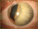
The Opportunities of Ultrasound Biomicroscopy in Diagnosis and Monitoring of Iridocorneal Endothelial Syndrome
The aim of the investigation is to estimate the opportunities of ultrasound biomicroscopy in diagnosis and monitoring in various types iridocorneal endothelial syndrome.
Materials and Methods. The study was carried out on the basis of a detailed analysis of scanning images: ultrasound eye biomicroscopy was performed on 29 patients with iridocorneal endothelial syndrome.
Results. There were revealed ultrasound microscopic features reflecting pathological changes in the anterior eye bulb.
Conclusion. The ultrasound microscopy value is that it enables to assess to the fullest extent the degree of anatomic structures involvement into pathological process as it is the only way to obtain life-time high resolution images of eye bulb “latent” zones. Moreover, using the technique iridocorneal endothelial syndrome can be differentiated from other pathological states with similar clinical picture. The authors recommend using the method for diagnosis and monitoring of various types of iridocorneal endothelial syndrome.
- Scheie H.G., Yanoff M. Arch Ophthalmol 1975; 93(10): 378–379.
- Wilson M.C., Shields M.B. Arch Ophthalmol 1989; 107(10): 1465–1468.
- Laganowski H.C., Kerr Muir M.G., Hitchings R.A. Arch Ophthalmol 1992; 110: 346–350.


