Common Path Side Viewing Monolithic Ball Lens Probe for Optical Coherence Tomography
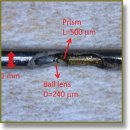
Common path probes are highly desirable for optical coherence tomography as they reduce system complexity and cost by eliminating the need of dispersion compensation and polarization controlling optics. In this work, we demonstrate a monolithic ball lens based, common path, side viewing probe that is suitable for Fourier domain optical coherence tomography. The probe design parameters were simulated in Zemax modeling software and the simulated performance parameters were compared with experimental results. We characterized the performance of the probe by measuring its collection efficiency, resolution, and sensitivity. Our results demonstrated that with a source input power of 25 mW, we could achieve sensitivity of 100.5 dB, which is within 7 dB of the maximum possible sensitivity that could be achieved using a separate reference arm. The axial resolution of the system was found to be 15.6 µm in air and the lateral resolution (full width half maximum) was approximately 49 µm. The probe optics were assembled in a 1 mm diameter hypotube with a 500 µm inner diameter. Images of finger skin acquired using this probe demonstrated clear visualization of the stratum corneum, epidermis, and papillary dermis, along with sweat ducts.
- Huang D., Swanson E.A., Lin C.P., Schuman J.S., Stinson W.G., Chang W., et al. Optical coherence tomography. Science 1991; 254(5035): 1178–1181, http://dx.doi.org/ 10.1126/science.1957169.
- Sergeev A.M., Gelikonov V.M., Gelikonov G.V., Feldchtein F.I., Kuranov R.V., Gladkova N.D., et al. In vivo endoscopic OCT imaging of precancer and cancer states of human mucosa. Opt Express 1997; 1(13): 432–440, http://dx.doi.org/10.1364/OE.1.000432.
- Wojtkowski M., Srinivasan V.J., Ko T.H., Fujimoto J.G., Kowalczyk A., Duker J.S. Ultrahigh-resolution, high-speed, Fourier domain optical coherence tomography and methods for dispersion compensation. Opt Express 2004; 12(11): 2404–2422, http://dx.doi.org/10.1364/OPEX.12.002404.
- Saxer C.E., de Boer J.F., Park B.H., Zhao Y., Chen Z., Nelson J.S. High-speed fiber based polarization-sensitive optical coherence tomography of in vivo human skin. Opt Lett 2000; 25(18): 1355–1357, http://dx.doi.org/10.1364/ol.25.001355.
- Zhang K., Kang J.U. Common-path low-coherence interferometry fiber-optic sensor guided microincision. J Biomed Opt 2011; 16(9): 095003, http://dx.doi.org/10.1117/1.3622492.
- Sharma U., Kang J.U. Common-path optical coherence tomography with side-viewing bare fiber probe for endoscopic optical coherence tomography. Rev Sci Instrum 2007; 78(11): 113102, http://dx.doi.org/10.1063/1.2804112.
- Han J.-H., Ilev I.K., Kim D.-H., Song C.G., Kang J.U. Investigation of gold-coated bare fiber probe for in situ intra-vitreous coherence domain optical imaging and sensing. Appl Phys B 2010; 99(4): 741–746, http://dx.doi.org/10.1007/s00340-010-3910-4.
- Ji-hyun Kim, Jae-Ho Han, Jichai Jeong. Common-path optical coherence tomography using a conical-frustum-tip fiber probe. IEEE J Select Topics Quantum Electron 2014; 20(2): 8–14, http://dx.doi.org/10.1109/jstqe.2013.2277817.
- Zhao M., Huang Y., Kang J.U. Sapphire ball lens-based fiber probe for common-path optical coherence tomography and its applications in corneal and retinal imaging. Opt Lett 2012; 37(23): 4835–4837, http://dx.doi.org/10.1364/OL.37.004835.
- Gelikonov V.M., Gelikonov G.V. New approach to cross-polarized optical coherence tomography based on orthogonal arbitrarily polarized modes. Laser Phys Lett 2006; 3(9): 445–451, http://dx.doi.org/10.1002/lapl.200610030.
- Lorenser D., Quirk B.C., Auger M., Madore W.-J., Kirk R.W., Godbout N., et al. Dual-modality needle probe for combined fluorescence imaging and three-dimensional optical coherence tomography. Opt Lett 2013; 38(3): 266–268, http://dx.doi.org/10.1364/OL.38.000266.
- Mao Y., Chang S., Sherif S., Flueraru C. Graded-index fiber lens proposed for ultrasmall probes used in biomedical imaging. Appl Opt 2007; 46(23): 5887–5894, http://dx.doi.org/10.1364/AO.46.005887.
- Yang V.X.D, Mao Y.X., Munce N., Standish B., Kucharczyk W., Marcon N.E., Vitkin I.A. Interstitial Doppler optical coherence tomography. Opt Lett 2005; 30(14): 1791–1793, http://dx.doi.org/10.1364/OL.30.001791.
- Leitgeb R., Hitzenberger C., Fercher A. Performance of fourier domain vs. time domain optical coherence tomography. Opt Express 2003; 11(8): 889–894, http://dx.doi.org/10.1364/OE.11.000889.


