Automated Method to Analyze Geometry and Topology of Mitral Valve Fibrous Ring
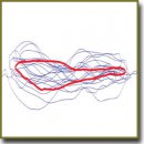
The design of prosthetic rings for the annuloplasty of mitral valve fibrous ring (MVFR) was developed from the intuitive to that based on the theoretical analysis of the geometry and topology using the modern technologies of image processing and analysis. The paper presents an automated method of three-dimensional echocardiography data processing and analysis followed by findings fitting.The technique includes several steps. At first, we detected the boundaries of the data obtained by transesophageal three-dimensional echocardiography followed by Procrustes analysis and the mean shape search based on the available data bulk. The method enables to analyze better MVFR geometry, develop MVFR models on the basis of the obtained data, and subsequently, use them to design a prosthetic ring in order to reduce the number of possible complications. Three approximation methods (piecewise cubic, general polynomial, Gauss–Fourier method) were applied for the optimization of the technique results. Each of the methods has its own unique set of advantages and disadvantages, and depending on the task, one can use a certain technique when analyzing or reconstructing MVFR geometry. Thus, it is a powerful tool for MVFR study and prosthetic ring modeling.
- Andreas M., Doll N., Livesey S., Castella M., Kocher A., Casselman F., Voth V., Bannister C., Encalada Palacios J.F., Pereda D., Laufer G., Czesla M. Safety and feasibility of a novel adjustable mitral annuloplasty ring: a multicentre European experience. Eur J Cardiothorac Surg 2016; 49(1): 249–254, http://dx.doi.org/10.1093/ejcts/ezv015.
- Carpentier A. Reconstructive valvuloplasty. A new technique of mitral valvuloplasty. Presse Med 1969; 77(7): 251–253.
- Lee L.S., Kwon M.H., Cevasco M., Schmitto J.D., Mokashi S.A., McGurk S., Cohn L.H., Bolman R.M. 3rd, Chen F.Y. Postoperative recurrence of mitral regurgitation after annuloplasty for functional mitral regurgitation. Ann Thorac Surg 2012; 94(4): 1211–1216, http://dx.doi.org/10.1016/j.athoracsur.2012.05.005.
- García E., Sandoval J., Unzue L., Hernandez-Antolin R., Almería C., Macaya C. Paravalvular leaks: mechanisms, diagnosis and management. EuroIntervention 2012; 8(Suppl Q): Q41–Q52, http://dx.doi.org/10.4244/EIJV8SQA9.
- Tsuneto A., Eishi K., Miura T., Tanigawa K., Matsukuma S., Minami T., Koide Y., Ikeda S., Kawano H., Maemura K. Comparison of saddle-shape flexibility and elliptical-shape stability between Cosgrove-Edwards and Memo-3D annuloplasty rings using three-dimensional analysis software. Gen Thorac Cardiovasc Surg 2016 Apr 6, http://dx.doi.org/10.1007/s11748-016-0645-0. [Epub ahead of print].
- Carpentier A. Mitral valve annuloplasty. Ann Thorac Surg 1990; 49(3): 508–509, http://dx.doi.org/10.1016/0003-4975(90)90277-d.
- Owais K., Montealegre-Gallegos M., Jeganathan J., Matyal R., Khabbaz K.R., Mahmood F. Dynamic changes in the ischemic mitral annulus: implications for ring sizing. Ann Card Anaesth 2016; 19(1): 15–19, http://dx.doi.org/10.4103/0971-9784.173014.
- Bayramoğlu A., Taşolar H., Otlu Y.Ö., Hidayet Ş., Kurt F., Doğan A., Pekdemir H. Assessment of left atrial volume and mechanical functions using real-time three-dimensional echocardiography in patients with mitral annular calcification. Anatol J Cardiol 2016; 16(1): 42–47, http://dx.doi.org/10.5152/akd.2015.5897.
- Salgo I.S., Gorman J.H. 3rd, Gorman R.C., Jackson B.M., Bowen F.W., Plappert T., St John Sutton M.G., Edmunds L.H. Jr. Effect of annular shape on leaflet curvature in reducing mitral leaflet stress. Circulation 2003; 106(6): 711–717, http://dx.doi.org/10.1161/01.cir.0000025426.39426.83.
- Pouch A.M., Vergnat M., McGarvey J.R., Ferrari G., Jackson B.M., Sehgal C.M., Yushkevich P.A., Gorman R.C., Gorman J.H. 3rd. Statistical assessment of normal mitral annular geometry using automated 3D echocardiographic analysis. Ann Thorac Surg 2014; 97(1): 71–77, http://dx.doi.org/10.1016/j.athoracsur.2013.07.096.


