EEG Correlates of Tactile Perception Abnormalities in Children with Autism Spectrum Disorder
The aim of the investigation was to study the changes in EEG power and behavioral responses to C-tactile stimulation in typically developing (TD) children and children with autism spectrum disorder (ASD).
Materials and Methods. EEG to manually delivered tactile stimuli was recorded for 79 children (ASD=39, TD=40) aged 5 to 10 years. CARS scores were obtained for each participant immediately before the recording session. The study involved recording resting EEG in eyes open condition within 1–2 min and collecting EEG response to tactile stimuli delivered pseudo-randomly for 3 experimental conditions (stroking with a soft brush, stroking with a harsh brush, and stimulation with a spiked roller delivered to the outer side of right forearm, stroking velocity was within 2–5 cm/s). Behavioral responses obtained by video recording during the experiment were assessed and coded. Behavioral responses were classified into 5 patterns: 1) signs of relaxation (facial gesture and body posture); 2) signs of resistance, attempts to withdraw the hand; 3) negative emotions, crying, shouting; 4) positive emotions, smile, laughter; 5) looking at the hand being stimulated. EEG power in 18 narrow frequency bands with a bandwidth of 1 Hz in a range of 2–20 Hz was analyzed.
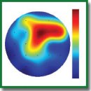
Results. The study revealed two types of response to tactile stimulation. The first type was not specific for particular tactile stimulation type, was accompanied by an increase in beta power (16–20 Hz) mainly in the left hemisphere and was more common in children with ASD. The second type of response was accompanied by an increase in frontal theta power (4–6 Hz) due to C-tactile system stimulation with a soft brush and was observed only in the TD children. The first type of response was accompanied by negative emotions and attempts to withdraw the hand, while the second type was characterized by relaxation.
Conclusion. The response of children with ASD to all types of tactile stimulation accompanied by an increase in beta power can be associated with both hypersensitivity and stress reaction of these children to the experimental situation. Selective response to C-tactile stimulation accompanied by an increase in frontal theta power has been found in the control group (TD) only. The results of this study can be useful for better understanding of hypersensitivity in children with ASD and gaining insight into the mechanisms of the disease.
- DiCicco-Bloom E., Lord C., Zwaigenbaum L., Courchesne E., Dager S.R., Schmitz C., Schultz R.T., Crawley J., Young L.J. The developmental neurobiology of autism spectrum disorder. J Neurosci 2006; 26(26): 6897–6906, https://doi.org/10.1523/jneurosci.1712-06.2006.
- Tomchek S.D., Dunn W. Sensory processing in children with and without autism: a comparative study using the short sensory profile. Am J Occup Ther 2007; 61(2): 190–200, https://doi.org/10.5014/ajot.61.2.190.
- Marco E.J., Hinkley L.B.N., Hill S.S., Nagarajan S.S. Sensory processing in autism: a review of neurophysiologic findings. Pediatric Research 2011; 69(5 Part 2): 48R–54R, https://doi.org/10.1203/PDR.0b013e3182130c54.
- Kanner L. Problems of nosology and psychodynamics of early infantile autism. Am J Orthopsychiatry 1949; 19(3): 416–426, https://doi.org/10.1111/j.1939-0025.1949.tb05441.x.
- Johansson R.S., Trulsson M., Olsson K.Å., Westberg K.-G. Mechanoreceptor activity from the human face and oral mucosa. Exp Brain Res 1988; 72(1): 204–208, https://doi.org/10.1007/bf00248518.
- McGlone F., Wessberg J., Olausson H. Discriminative and
affective touch: sensing and feeling. Neuron 2014; 82(4): 737–755, https://doi.org/10.1016/j.neuron.2014.05.001. - Baranek G.T. Autism during infancy: a retrospective video analysis of sensory-motor and social behaviors at 9–12 months of age. J Autism Dev Disord 1999; 29(3): 213–224, https://doi.org/10.1023/a:1023080005650.
- Yang D.Y., Rosenblau G., Keifer C., Pelphrey K.A. An integrative neural model of social perception, action observation, and theory of mind. Neurosci Biobehav Rev 2015; 51(8): 263–275, https://doi.org/10.1016/j.neubiorev.2015.01.020.
- Kaiser M.D., Yang D.Y., Voos A.C., Bennett R.H., Gordon I., Pretzsch C., Beam D., Keifer C., Eilbott J., McGlone F., Pelphrey K.A. Brain mechanisms for processing affective (and nonaffective) touch are atypical in autism. Cereb Cortex 2016; 26(6): 2705–2714, https://doi.org/10.1093/cercor/bhv125.
- Bennett R.H., Bolling D.Z., Anderson L.C., Pelphrey K.A., Kaiser M.D. fNIRS detects temporal lobe response to
affective touch. Soc Cogn Affect Neurosci 2014; 9(4): 470–476, https://doi.org/10.1093/scan/nst008. - Löken L.S., Wessberg J., Morrison I., McGlone F., Olausson H. Coding of pleasant touch by unmyelinated afferents in humans. Nat Neurosci 2009; 12(5): 547–548, https://doi.org/10.1038/nn.2312.
- Schopler E., Reichler R.J., DeVellis R.F., Daly K. Toward objective classification of childhood autism: Childhood Autism Rating Scale (CARS). J Autism Dev Disord 1980; 10(1): 91–103, https://doi.org/10.1007/bf02408436.
- Wechsler D. Weschler Intelligence Scale for Children: third edition manual. San Antonio, TX: The Psychological Corporation; 1991.
- Klem G.H., Lüders H.O., Jasper H.H., Elger C. The ten-twenty electrode system of the International Federation. The International Federation of Clinical Neurophysiology. Electroencephalogr Clin Neurophysiol Suppl 1999; 52: 3–6.
- Murias M., Webb S.J., Greenson J., Dawson G. Resting state cortical connectivity reflected in EEG coherence in individuals with autism. Biol Psychiatry 2007; 62(3): 270–273, https://doi.org/10.1016/j.biopsych.2006.11.012.
- Gasser T., Verleger R., Bächer P., Sroka L. Development of the EEG of school-age children and adolescents. I. Analysis of band power. Electroencephalogr Clin Neurophysiol 1988; 69(29): 91–99, https://doi.org/10.1016/0013-4694(88)90204-0.
- Fraga González G., Van der Molen M.J.W., Žarić G., Bonte M., Tijms J., Blomert L., Stam C.J., Van der Molen M.W. Graph analysis of EEG resting state functional networks in dyslexic readers. Clin Neurophysiol 2016; 127(9): 3165–3175, https://doi.org/10.1016/j.clinph.2016.06.023.
- Szurhaj W., Derambure P., Labyt E., Cassim F., Bourriez J.L., Isnard J., Guieu J.D., Mauguière F. Basic mechanisms of central rhythms reactivity to preparation and execution of a voluntary movement: a stereoelectroencephalographic study. Clin Neurophysiol 2003; 114(1): 107–119, https://doi.org/10.1016/s1388-2457(02)00333-4.
- Ohara S., Ikeda A., Kunieda T., Yazawa S., Baba K., Nagamine T., Taki W., Hashimoto N., Mihara T., Shibasaki H. Movement-related change of electrocorticographic activity in human supplementary motor area proper. Brain 2000; 123(6): 1203–1215, https://doi.org/10.1093/brain/123.6.1203.
- Güntekin B., Başar E. Event-related beta oscillations are affected by emotional eliciting stimuli. Neurosci Lett 2010; 483(3): 173–178, https://doi.org/10.1016/j.neulet.2010.08.002.
- Roohi-Azizi M., Azimi L., Heysieattalab S.,
Aamidfar M. Changes of the brain’s bioelectrical activity in cognition, consciousness, and some mental disorders. Med J Islam Repub Iran 2017; 31(1): 307–312, https://doi.org/10.14196/mjiri.31.53. - Cowan J., Markham L. EEG biofeedback for the attention problems of autism: a case study. In: 25th annual meeting of the Association for Applied Psychophysiology and Biofeedback. 1994; p. 12–13.
- Cascio C.J., Moana-Filho E.J., Guest S., Nebel M.B., Weisner J., Baranek G.T., Essick G.K. Perceptual and neural response to affective tactile texture stimulation in adults with autism spectrum disorders. Autism Res 2012; 5(4): 231–244, https://doi.org/10.1002/aur.1224.
- Cascio C.J. Somatosensory processing in neurodevelopmental disorders. J Neurodev Disord 2010; 2(2): 62–69, https://doi.org/10.1007/s11689-010-9046-3.
- Guclu B., Tanidir C., Mukaddes N.M. Tactile sensitivity of normal and autistic children. Somatosens Mot Res 2007; 24(1–2): 21–33, https://doi.org/10.1080/08990220601179418.
- Singh H., Bauer M., Chowanski W., Sui Y., Atkinson D., Baurley S., Fry M., Evans J., Bianchi-Berthouze N. The brain’s response to pleasant touch: an EEG investigation of tactile caressing. Front Hum Neurosci 2014; 8: 893, https://doi.org/10.3389/fnhum.2014.00893.
- von Mohr M., Crowley M.J., Walthall J., Mayes L.C., Pelphrey K.A., Rutherford H.J.V. EEG captures affective touch: CT-optimal touch and neural oscillations. Cogn Affect Behav Neurosci 2018; 18(1): 155–166, https://doi.org/10.3758/s13415-017-0560-6.
- Ackerley R., Eriksson E., Wessberg J. Ultra-late EEG potential evoked by preferential activation of unmyelinated tactile afferents in human hairy skin. Neurosci Lett 2013; 535: 62–66, https://doi.org/10.1016/j.neulet.2013.01.004.
- Crane L., Goddard L., Pring L. Sensory processing in adults with autism spectrum disorders. Autism 2009; 13(3): 215–228, https://doi.org/10.1177/1362361309103794.
- Björnsdotter M., Gordon I., Pelphrey K.A., Olausson H., Kaiser M.D. Development of brain mechanisms for processing affective touch. Front Behav Neurosci 2014; 8: 24, https://doi.org/10.3389/fnbeh.2014.00024.


