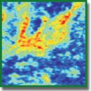
Contrast Enhancement of Cross-Polarization OCT Images of Breast Cancer by Optical Coefficient Calculation
The aim of the study was to develop the techniques for contrast enhancement of cross-polarization OCT (CP OCT) images of breast cancer (BC) by calculating optical attenuation coefficients of OCT signal to differentiate cancer subtypes and analyze polarization properties of tumor stroma.
Materials and Methods. The study involved ex vivo breast tissue samples after radical mastectomy. Scattering and polarization properties were studied using a high-speed spectral OCT unit suitable for cross-scattering recording with higher specificity enabling to differentiate breast connective tissue in health and cancer. We presented the findings of qualitative and quantitative analysis of en face CP OCT images of non-tumor breast cancer and two the most common BC types, which differ fundamentally by tumor stroma condition — infiltrating ductal carcinoma of solid and sclerosing structures. Optical coefficients were calculated for contrast enhancement of CP OCT images: attenuation coefficient in a co-channel (coefficient 1) and a difference attenuation coefficient in co- and cross-channels (coefficient 2), which enabled both: to differ tumor tissue from non-tumor tissue, and differentiate two breast cancer types under study.
Results. En face CP OCT images of breast tissue without tumor are characterized by heterogeneity due to different scattering capacity of fat, glandular and connective tissue with predominantly low values of both optical coefficients. Infiltrating ductal BC of a solid structure is characterized by homogenous distribution of lowest values of both optical coefficients. In case of infiltrating ductal BC of a sclerosing structure, CP OCT images have the most heterogeneous and contrast OCT signal related to the tumor stroma dominated over parenchyma, and various degenerative changes are in stroma (fibrosis or hyalinosis).
Conclusion. The use of optical attenuation coefficients of OCT signal in two channels and en face color-coded mapping is an obvious example presenting CP OCT images of BC and an objective technique for OCT signal quantitative assessment. Moreover, imaging contrast enhances, it eases the differentiation of morphological BC subtypes and can be used in clinical settings.
- https://www.cancer.org/research/cancer- facts-statistics/breast-cancer-facts-figures.html.
- https://media.nenaprasno.ru/cancer- catalog/additional-articles/1652/.
- Zlokachestvennye novoobrazovaniya v Rossii v 2017 godu (zabolevaemost’ i smertnost’) [Malignant neoplasms in Russia in 2017 (morbidity and mortality)]. Pod red. Kaprina A.D., Starinskogo V.V., Petrovoy G.V. [Kaprin A.D., Starinskiy V.V., Petrova G.V. (editors)]. Moscow: MNIOI im. P.A. Gertsena — filial FGBU “NMITs radiologii” Minzdrava Rossii; 2018; 250 p.
- Aleskandarany M.A., Vandenberghe M.E., Marchiò C., Ellis I.O., Sapino A., Rakha E.A. Tumour heterogeneity of breast cancer: from morphology to personalised medicine. Pathobiology 2018; 85(1–2): 23–34, https://doi.org/10.1159/000477851.
- WHO classification of tumours of the breast. Edited by Lakhani S.R., Ellis I.O., Schnitt S.J., Tan P.H., van de Vijver M.J. International Agency for Research on Cancer; 2012.
- Cèfaro G.A., Genovesi D., Marchese R., Ursini L.A., Cianchetti E., Ballone E., Di Nicola M. Predictors of local recurrence after conservative surgery and whole-breast irradiation. Breast Cancer Res Treat 2006; 98(3): 329–335, https://doi.org/10.1007/s10549-006-9169-0.
- Luini A., Rososchansky J., Gatti G., Zurrida S., Caldarella P., Viale G., Rosali dos Santos G., Frasson A. The surgical margin status after breast-conserving surgery: discussion of an open issue. Breast Cancer Res Treat 2008; 113(2): 397–402, https://doi.org/10.1007/s10549-008-9929-0.
- Catalano O.A., Horn G.L., Signore A., Iannace C., Lepore M., Vangel M., Luongo A., Catalano M., Lehman C., Salvatore M., Soricelli A., Catana C., Mahmood U., Rosen B.R. PET/MR in invasive ductal breast cancer: correlation between imaging markers and histological phenotype. Br J Cancer 2017; 116(7): 893–902, https://doi.org/10.1038/bjc.2017.26.
- Kuhl C.K. Abbreviated magnetic resonance imaging (MRI) for breast cancer screening: rationale, concept, and transfer to clinical practice. Annu Rev Med 2019; 70(1): 501–519, https://doi.org/10.1146/annurev-med-121417-100403.
- Taylor K.J., Merritt C., Piccoli C., Schmidt R., Rouse G., Fornage B., Rubin E., Georgian-Smith D., Winsberg F., Goldberg B., Mendelson E. Ultrasound as a complement to mammography and breast examination to characterize breast masses. Ultrasound Med Biol 2002; 28(1): 19–26, https://doi.org/10.1016/s0301-5629(01)00491-4.
- Evans A., Trimboli R.M., Athanasiou A., Balleyguier C., Baltzer P.A., Bick U., Camps Herrero J., Clauser P., Colin C., Cornford E., Fallenberg E.M., Fuchsjaeger M.H., Gilbert F.J., Helbich T.H., Kinkel K., Heywang-Köbrunner S.H., Kuhl C.K., Mann R.M., Martincich L., Panizza P., Pediconi F., Pijnappel R.M., Pinker K., Zackrisson S., Forrai G., Sardanelli F.; European Society of Breast Imaging (EUSOBI), with language review by Europa Donna–The European Breast Cancer Coalition. Breast ultrasound: recommendations for information to women and referring physicians by the European Society of Breast Imaging. Insights Imaging 2018; 9(4): 449–461, https://doi.org/10.1007/s13244-018-0636-z.
- Esbona K., Li Z., Wilke L.G. Intraoperative imprint cytology and frozen section pathology for margin assessment in breast conservation surgery: a systematic review. Ann Surg Oncol 2012; 19(10): 3236–3245, https://doi.org/10.1245/s10434-012-2492-2.
- Yao X., Gan Y., Chang E., Hibshoosh H., Feldman S., Hendon C. Visualization and tissue classification of human breast cancer images using ultrahigh-resolution OCT. Lasers Surg Med 2017; 49(3): 258–269, https://doi.org/10.1002/lsm.22654.
- Zhou C., Cohen D.W., Wang Y., Lee H.C., Mondelblatt A.E., Tsai T.H., Aguirre A.D., Fujimoto J.G., Connolly J.L. Integrated optical coherence tomography and microscopy for ex vivo multiscale evaluation of human breast tissues. Cancer Res 2010; 70(24): 10071–10079, https://doi.org/10.1158/0008-5472.can-10-2968.
- Ha R., Friedlander L.C., Hibshoosh H., Hendon C., Feldman S., Ahn S., Schmidt H., Akens M.K., Fitzmaurice M., Wilson B.C., Mango V.L. Optical coherence tomography: a novel imaging method for post-lumpectomy breast margin assessment-a multi-reader study. Acad Radiol 2018; 25(3): 279–287, https://doi.org/10.1016/j.acra.2017.09.018.
- Nguyen F.T., Zysk A.M., Chaney E.J., Kotynek J.G., Oliphant U.J., Bellafiore F.J., Rowland K.M., Johnson P.A., Boppart S.A. Intraoperative evaluation of breast tumor margins with optical coherence tomography. Cancer Research 2009; 69(22): 8790–8796, https://doi.org/10.1158/0008-5472.can-08-4340.
- Savastru D., Chang E.W., Miclos S., Pitman M.B., Patel A., Iftimia N. Detection of breast surgical margins with optical coherence tomography imaging: a concept evaluation study. J Biomed Opt 2014; 19(5): 056001, https://doi.org/10.1117/1.jbo.19.5.056001.
- Mujat M., Ferguson R.D., Hammer D.X., Gittins C., Iftimia N. Automated algorithm for breast tissue differentiation in optical coherence tomography. J Biomed Opt 2009; 14(3): 034040, https://doi.org/10.1117/1.3156821.
- Zysk A.M., Boppart S.A. Computational methods for analysis of human breast tumor tissue in optical coherence tomography images. J Biomed Opt 2006; 11(5): 054015, https://doi.org/10.1117/1.2358964.
- Zysk A.M., Chaney E.J., Boppart S.A. Refractive index of carcinogen-induced rat mammary tumours. Phys Med Biol 2006; 51(9): 2165–2177, https://doi.org/10.1088/0031-9155/51/9/003.
- Hsiung P.-L., Phatak D.R., Chen Y., Aguirre A.D., Fujimoto J.G., Connolly J.L. Benign and malignant lesions in the human breast depicted with ultrahigh resolution and three-dimensional optical coherence tomography. Radiology 2007; 244(3): 865–874, https://doi.org/10.1148/radiol.2443061536.
- South F.A., Chaney E.J., Marjanovic M., Adie S.G., Boppart S.A. Differentiation of ex vivo human breast tissue using polarization-sensitive optical coherence tomography. Biomed Opt Express 2014; 5(10): 3417, https://doi.org/10.1364/boe.5.003417.
- Patel R., Khan A., Quinlan R., Yaroslavsky A.N. Polarization-sensitive multimodal imaging for detecting breast cancer. Cancer Res 2014; 74(17): 4685–4693, https://doi.org/10.1158/0008-5472.can-13-2411.
- Chin L., Latham B., Saunders C.M., Sampson D.D., Kennedy B.F. Simplifying the assessment of human breast cancer by mapping a micro-scale heterogeneity index in optical coherence elastography. J Biophotonics 2016; 10(5): 690–700, https://doi.org/10.1002/jbio.201600092.
- Kennedy B.F., McLaughlin R.A., Kennedy K.M., Chin L., Wijesinghe P., Curatolo A., Tien A., Ronald M., Latham B., Saunders C.M., Sampson D.D. Investigation of optical coherence microelastography as a method to visualize cancers in human breast tissue. Cancer Res 2015; 75(16): 3236–3245, https://doi.org/10.1158/0008-5472.can-14-3694.
- Gubarkova E.V., Sovetsky A.A., Zaitsev V.Y., Matveyev A.L., Vorontsov D.A., Sirotkina M.A., Matveev L.A., Plekhanov A.A., Pavlova N.P., Kuznetsov S.S., Vorontsov A.Y., Zagaynova E.V., Gladkova N.D. OCT-elastography-based optical biopsy for breast cancer delineation and express assessment of morphological/molecular subtypes. Biomed Opt Express 2019; 10(5): 2244, https://doi.org/10.1364/boe.10.002244.
- Gelikonov V.M., Romashov V.N., Shabanov D.V., Ksenofontov S.Y., Terpelov D.A., Shilyagin P.A., Gelikonov G.V., Vitkin I.A. Cross-polarization optical coherence tomography with active maintenance of the circular polarization of a sounding wave in a common path system. Radiophys Quantum El 2018; 60(11): 897–911, https://doi.org/10.1007/s11141-018-9856-9.
- Gubarkova E.V., Dudenkova V.V., Feldchtein F.I., Timofeeva L.B., Kiseleva E.B., Kuznetsov S.S., Shakhov B.E., Moiseev A.A., Gelikonov V.M., Gelikonov G.V., Vitkin A., Gladkova N.D. Multi-modal optical imaging characterization of atherosclerotic plaques. J Biophotonics 2015; 9(10): 1009–1020, https://doi.org/10.1002/jbio.201500223.
- Kiseleva E., Kirillin M., Feldchtein F., Vitkin A., Sergeeva E., Zagaynova E., Streltzova O., Shakhov B., Gubarkova E., Gladkova N. Differential diagnosis of human bladder mucosa pathologies in vivo with cross-polarization optical coherence tomography. Biomed Opt Express 2015; 6(4): 1464–1476, https://doi.org/10.1364/boe.6.001464.
- Rak molochnoy zhelezy. Morfologicheskaya diagnostika i genetika [Breast cancer. Morphological diagnosis and genetics]. Pod red. Franka G.A., Zavalishinskoy L.E., Pozharisskogo K.M. [Frank G.A., Zavalishinskaya L.E., Pozharisskiy K.M. (editors)]. Moscow: Prakticheskaya meditsina; 2014.
- Kut C., Chaichana K.L., Xi J., Raza S.M., Ye X., McVeigh E.R., Rodriguez F.J., Quiñones-Hinojosa A., Li X. Detection of human brain cancer infiltration ex vivo and in vivo using quantitative optical coherence tomography. Sci Transl Med 2015; 7(292): 3010611, https://doi.org/10.1126/scitranslmed.3010611.
- Provenzano P.P., Eliceiri K.W., Campbell J.M., Inman D.R., White J.G., Keely P.J. Collagen reorganization at the tumor-stromal interface facilitates local invasion. BMC Med 2006; 4(1): 38, https://doi.org/10.1186/1741-7015-4-38.


