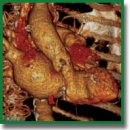
A New Look at Structural Changes in the Aortic Root in Aortic Valve Stenosis
The aim of the study was to identify new anatomical landmarks of the aortic root and the relationship between the sizes of anatomical structures using the method of computed tomography angiography to improve models of heart valves and the methods for their selection in clinical practice.
Materials and Methods. The dataset of computed tomography angiography prior to aortic valve replacement in 262 patients was analyzed. The mean age was 75.0±5.9 years. 99 (37.8±3.0%) men and 163 (62.2±3.0%) women took part in the study. The annulus fibrosus, sinotubular junction, and height of the sinuses of Valsalva were measured.
Results. In the tricuspid aortic valve group (n=251), in more than 50% of the cases, the diameter of the annulus fibrosus ranged from 23 to 26 mm. No significant association between the diameter of the annulus fibrosus and patient height (r=0.35; p=0.01) or body surface area (r=0.25; p=0.01) and the height of the sinuses of Valsalva (r=0.34; p=0.01) were revealed. Based on the ratio of the height of the sinuses of Valsalva and the diameter of the annulus fibrosus, three variants of the structure of the aortic root were identified: type A — K>1.05; type B — 0.95≤K≤1.05; type C — K<0.95. Type C of the aortic root was found to predominate in most cases, namely, in 98.0±0.9% (n=246).
In the bicuspid aortic valve group (n=11), 2 patients had a type A of the aortic root, 1 patient had a type B, and 8 patients had a type C.
Conclusion. A classification of variants of the aortic root structure has been proposed, which will be useful not only for practitioners when choosing a treatment method, but also for researchers to understand the structural characteristics of the aortic root in patients with its pathology.
- Bonow R.O., Greenland P. Population-wide trends in aortic stenosis incidence and outcomes. Circulation 2015; 131(11): 969–971, https://doi.org/10.1161/circulationaha.115.014846.
- Eggebrecht H., Mehta R.H. Transcatheter aortic valve implantation (TAVI) in Germany 2008–2014: on its way to standard therapy for aortic valve stenosis in the elderly? EuroIntervention 2016; 11(9): 1029–1033, https://doi.org/10.4244/eijy15m09_11.
- Tchetche D., Van Mieghem N.M. New-generation TAVI devices: description and specifications. EuroIntervention 2014; 10(Suppl U): U90–U100, https://doi.org/10.4244/eijv10sua13.
- Randhawa A., Gupta T., Singh P., Aggarwal A., Sahni D. Description of the aortic root anatomy in relation to transcatheter aortic valve implantation. Cardiovasc Pathol 2019; 40: 19–23, https://doi.org/10.1016/j.carpath.2019.01.005.
- Sud A., Parker F., Magilligan D.J. Jr. Anatomy of the aortic root. Ann Thorac Surg 1984; 38(1): 76–79, https://doi.org/10.1016/s0003-4975(10)62195-9.
- Ait Said M., Coquard C., Horvilleur J., Manenti V., Fiorina L., Lacotte J., Salerno F. Transcatheter aortic valve implantation and conduction disturbances. Ann Cardiol Angeiol (Paris) 2019; 68(6): 443–449, https://doi.org/10.1016/j.ancard.2019.09.024.
- Achenbach S., Delgado V., Hausleiter J., Schoenhagen P., Min J.K., Leipsic J.A. SCCT expert consensus document on computed tomography imaging before transcatheter aortic valve implantation (TAVI)/transcatheter aortic valve replacement (TAVR). J Cardiovasc Comput Tomogr 2012; 6(6): 366–380, https://doi.org/10.1016/j.jcct.2012.11.002.
- Du Bois D., Du Bois E.F. A formula to estimate the approximate surface area if height and weight be known. 1916. Nutrition 1989; 5(5): 303–313.
- Bokeriya L.A., Skopin I.I., Sazonenkov M.A., Tumaev E.N. On the question of the anatomy of the aortic root. The ratio of the diameters of the aortic ring and the sinotubular junction is normal in adults. Ideal geometric model of the aortic root. Byulleten’ NTsSSKh im. A.N. Bakuleva RAMN 2008; 9(4): 77–85.
- Sophocleous F., Berlot B., Ordonez M.V., Baquedano M., Milano E.G., De Francesco V., Stuart G., Caputo M., Bucciarelli-Ducci C., Biglino G. Determinants of aortic growth rate in patients with bicuspid aortic valve by cardiovascular magnetic resonance. Open Heart 2019; 6(2): e001095, https://doi.org/10.1136/openhrt-2019-001095.
- Vincent F., Ternacle J., Denimal T., Shen M., Redfors B., Delhaye C., Simonato M., Debry N., Verdier B., Shahim B., Pamart T., Spillemaeker H., Schurtz G., Pontana F., Thourani V.H., Pibarot P., Van Belle E. Transcatheter aortic valve replacement in bicuspid aortic valve stenosis. Circulation 2021; 143(10): 1043–1061, https://doi.org/10.1161/circulationaha.120.048048.
- Ram D., Bouhout I., Karliova I., Schneider U., El-Hamamsy I., Schäfers H.J. Concepts of bicuspid aortic valve repair: a review. Ann Thorac Surg 2020; 109(4): 999–1006, https://doi.org/10.1016/j.athoracsur.2019.09.019.
- Bayandin N.L., Krotovsky A.G., Vasilyev K.N., Moiseev A.A., Setyn T.V. Comparative results of surgical and transcatheter (TAVI) treatment of aortic stenosis in patients over 75 years old. Rossijskij kardiologiceskij zurnal 2018; 11: 21–26, https://doi.org/10.15829/1560-4071-2018-11-21-26.
- Baumgartner H., Hung J., Bermejo J., Chambers J.B., Edvardsen T., Goldstein S., Lancellotti P., LeFevre M., Miller F. Jr., Otto C.M. Recommendations on the echocardiographic assessment of aortic valve stenosis: a focused update from the European Association of Cardiovascular Imaging and the American Society of Echocardiography. J Am Soc Echocardiogr 2017; 30(4): 372–392, https://doi.org/10.1016/j.echo.2017.02.009.
- Haj-Ali R., Marom G., Ben Zekry S., Rosenfeld M., Raanani E. A general three-dimensional parametric geometry of the native aortic valve and root for biomechanical modeling. J Biomech 2012; 45(14): 2392–2397, https://doi.org/10.1016/j.jbiomech.2012.07.017.
- Stukalova O.V., Serova N.S., Chepovskiy A.M., Ternovoy S.K. MRI-based computer modeling of the heart: clinical application in arrhythmology. Rossijskij elektronnyj zurnal lucevoj diagnostiki 2021; 11(2): 32–45.
- Hussein N., Voyer-Nguyen P., Portnoy S., Peel B., Schrauben E., Macgowan C., Yoo S.J. Simulation of semilunar valve function: computer-aided design, 3D printing and flow assessment with MR. 3D Print Med 2020; 6(1): 2, https://doi.org/10.1186/s41205-020-0057-8.
- Marom G., Haj-Ali R., Rosenfeld M., Schäfers H.J., Raanani E. Aortic root numeric model: annulus diameter prediction of effective height and coaptation in post-aortic valve repair. J Thorac Cardiovasc Surg 2013; 145(2): 406–411.e1, https://doi.org/10.1016/j.jtcvs.2012.01.080.


