Visualization of Gastric Adenocarcinoma Lymph Node Metastases by Microscopy with Ultraviolet Surface Excitation
The detection of lymph node metastases is crucial in oncopathology, as it makes it possible to determine the TNM stage, to design a treatment plan, and predict the survival for cancer patients. The current gold standard for this process is hematoxylin and eosin staining. However, new alternative methods leveraging the unique optical properties of tissue structures are being developed for rapid intraoperative or postoperative application.
The aim of the study is to evaluate the effectiveness of identifying lymph node metastases using microscopy with ultraviolet surface excitation (MUSE).
Materials and Methods. 17 lymph nodes from the Sechenov University archive (Russia) collected intraoperatively from 6 patients with gastric cancer have been investigated.
In this study, we utilized a MUSE optical system consisting of three UV light-emitting diodes (265 nm) and the Axio Scope A1 microscope (Carl Zeiss, Germany) with various objectives. We introduced a novel combination of fluorescent dyes — Nile red and Hoechst — that had not been previously used with MUSE.
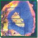
Results. The combination of fluorescent dyes yielded high-contrast images with blue-stained nuclei and orange-to-red stained cytoplasm, effectively visualizing gastric adenocarcinoma cells characterized by abundant cytoplasmic components and large polymorphic nuclei. The presence of irregularly shaped cavities, formed by adenocarcinoma metastases, was also detectable by MUSE.
Conclusion. Biophotonics provides alternative methods for tissue imaging. However, traditional methods are still unsurpassed in the accuracy of detecting cancer metastases and other pathologies. Further refinement of imaging protocols and expanded research into other cancer types are needed to make methods like MUSE applicable for intraoperative diagnosis.
- Feldman A.T., Wolfe D. Tissue processing and hematoxylin and eosin staining. Methods Mol Biol 2014; 1180: 31–43, https://doi.org/10.1007/978-1-4939-1050-2_3.
- TNM-atlas: illustrated guide to the TNM/pTNM-classification of malignant tumours. Spiessl B., Hermanek P., Scheibe O., Wagner G., Kerl U. (eds). Springer Science & Business Media; 2013.
- Mariette C., Piessen G., Briez N., Triboulet J.P. The number of metastatic lymph nodes and the ratio between metastatic and examined lymph nodes are independent prognostic factors in esophageal cancer regardless of neoadjuvant chemoradiation or lymphadenectomy extent. Ann Surg 2008; 247(2): 365–371, https://doi.org/10.1097/SLA.0b013e31815aaadf.
- Lawrence W.D.; Association of Directors of Anatomic and Surgical Pathology. ADASP recommendations for processing and reporting of lymph node specimens submitted for evaluation of metastatic disease. Virchows Arch 2001; 439(5): 601–603, https://doi.org/10.1007/s004280100412.
- Turner R.R., Ollila D.W., Krasne D.L., Giuliano A.E. Histopathologic validation of the sentinel lymph node hypothesis for breast carcinoma. Ann Surg 1997; 226(3): 271–278, https://doi.org/10.1097/00000658-199709000-00006.
- Yang H., Zhang S., Liu P., Cheng L., Tong F., Liu H., Wang S., Liu M., Wang C., Peng Y., Xie F., Zhou B., Cao Y., Guo J., Zhang Y., Ma Y., Shen D., Xi P., Wang S. Use of high-resolution full-field optical coherence tomography and dynamic cell imaging for rapid intraoperative diagnosis during breast cancer surgery. Cancer 2020; 126(Suppl 16): 3847–3856, https://doi.org/10.1002/cncr.32838.
- Tyszka J.M., Fraser S.E., Jacobs R.E. Magnetic resonance microscopy: recent advances and applications. Curr Opin Biotechnol 2005; 16(1): 93–99, https://doi.org/10.1016/j.copbio.2004.11.004.
- Kantere D., Siarov J., De Lara S., Parhizkar S., Olofsson Bagge R., Wennberg Larkö A., Ericson M.B. Label‐free laser scanning microscopy targeting sentinel lymph node diagnostics: a feasibility study ex vivo. Translational Biophotonics 2020; 2(3), https://doi.org/10.1002/tbio.202000002.
- Kong K., Kendall C., Stone N., Notingher I. Raman spectroscopy for medical diagnostics — from in-vitro biofluid assays to in-vivo cancer detection. Adv Drug Deliv Rev 2015; 89: 121–134, https://doi.org/10.1016/j.addr.2015.03.009.
- Sattlecker M., Bessant C., Smith J., Stone N. Investigation of support vector machines and Raman spectroscopy for lymph node diagnostics. Analyst 2010; 135(5): 895–901, https://doi.org/10.1039/b920229c.
- Fereidouni F., Harmany Z.T., Tian M., Todd A., Kintner J.A., McPherson J.D., Borowsky A.D., Bishop J., Lechpammer M., Demos S.G., Levenson R. Microscopy with ultraviolet surface excitation for rapid slide-free histology. Nat Biomed Eng 2017; 1(12): 957–966, https://doi.org/10.1038/s41551-017-0165-y.
- Liu Y., Rollins A.M., Levenson R.M., Fereidouni F., Jenkins M.W. Pocket MUSE: an affordable, versatile and high-performance fluorescence microscope using a smartphone. Commun Biol 2021; 4(1): 334, https://doi.org/10.1038/s42003-021-01860-5.
- Cooper D.J., Huang C., Klavins D.A., Fauver M.E., Carson M.D., Fereidouni F., Dintzis S., Galambos C., Levenson R.M., Seibel E.J. CoreView: fresh tissue biopsy assessment at the bedside using a millifluidic imaging chip. Lab Chip 2022; 22(7): 1354–1364, https://doi.org/10.1039/d1lc01142a.
- Kolluru C., Todd A., Upadhye A.R., Liu Y., Berezin M.Y., Fereidouni F., Levenson R.M., Wang Y., Shoffstall A.J., Jenkins M.W., Wilson D.L. Imaging peripheral nerve micro-anatomy with MUSE, 2D and 3D approaches. Sci Rep 2022; 12(1): 10205, https://doi.org/10.1038/s41598-022-14166-1.
- Yoshitake T., Giacomelli M.G., Quintana L.M., Vardeh H., Cahill L.C., Faulkner-Jones B.E., Connolly J.L., Do D., Fujimoto J.G. Rapid histopathological imaging of skin and breast cancer surgical specimens using immersion microscopy with ultraviolet surface excitation. Sci Rep 2018; 8(1): 4476, https://doi.org/10.1038/s41598-018-22264-2.
- Fereidouni F., Mitra A.D., Demos S., Levenson R. Microscopy with UV surface excitation (MUSE) for slide-free histology and pathology imaging. Optical Biopsy XIII: Toward Real-Time Spectroscopic Imaging and Diagnosis 2015; 9318: 46–51.
- Levenson R., Fereidouni F., Harmany Z., Tan M., Lechpammer M., Demos S. Slide-free microscopy via UV surface excitation. Microscopy and Microanalysis 2016; 22: 1002–1003, https://doi.org/10.1017/s1431927616005857.
- Xie W., Chen Y., Wang Y., Wei L., Yin C., Glaser A.K., Fauver M.E., Seibel E.J., Dintzis S.M., Vaughan J.C., Reder N.P., Liu J.T.C. Microscopy with ultraviolet surface excitation for wide-area pathology of breast surgical margins. J Biomed Opt 2019; 24(2): 1–11, https://doi.org/10.1117/1.JBO.24.2.026501.
- Matsumoto T., Niioka H., Kumamoto Y., Sato J., Inamori O., Nakao R., Harada Y., Konishi E., Otsuji E., Tanaka H., Miyake J., Takamatsu T. Deep-UV excitation fluorescence microscopy for detection of lymph node metastasis using deep neural network. Sci Rep 2019; 9(1): 16912, https://doi.org/10.1038/s41598-019-53405-w.
- Greenspan P., Fowler S.D. Spectrofluorometric studies of the lipid probe, nile red. J Lipid Res 1985; 26(7): 781–789.


