Diagnostic Ultrasound of Diffuse-Nodular Pleural Mesotheliomas — Echosemiotics, Growth Characteristics, and Scanning Techniques
The aim of the investigation was the optimization of chest ultrasound technique in the diagnosis of diffuse-nodular pleural mesotheliomas, development and systematization of their ultrasound semiotics.
Materials and Methods. We studied the ultrasonic picture of 32 mesotheliomas (31 malignant), among them 30 — diffuse-nodular, 2 — focal.
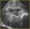
Results. There were developed the assessment criteria of diffuse-nodular mesothelioma echosemiotics: significant (over 15 mm) diffuse thickening of costal and diaphragmal pleura, with homogeneous hypoechoic structure and sharp contour against a marked (700–1500 ml) anechoic pleural effusion. For the first time we revealed the predominance of anterior costal and diaphragmal pleural lesions with typical caudal extension of the whole phrenicocostal sinus outwards. Chest ultrasonography optimization in mesothelioma diagnosis consists in longitudinal chest scanning from parasternal to anterior axillary line, first in V–VII intercostals space and then under costal arch with the following probe displacement down the anterior abdominal wall to assess the tumor extension in soft tissues. Typical deformity and diaphragm thinning resulted from tumor extension into diaphragm were described.
Conclusion. Chest ultrasonography is a high-quality diagnostic technique of diffuse-nodular mesothelioma enabling to determine tumor malignance, its extension, size, and invasion in surrounding tissues.
- Yablonskiy P.K., Petrov A.S. Malignant pleural mesothelioma. Prakticheskaya onkologiya 2006; 3: 179–188.
- Sinyukova G.T., Sholokhov V.N., Gudilina E.A. Diagnostic ultrasound of pleural masses. Ul’trazvukovaya diagnostika 2000; 1: 98–101.
- Beckh S., Blcskei P.L., Lessnau K.D. Real-time chest ultrasonography: a comprehensive review for the pulmonologist. Chest 2002; 122(5): 1759–1773, http://dx.doi.org/10.1378/chest.122.5.1759.
- Reuβ J. Sonographie der pleura. Ultraschall Med 2010; 31(1): 8–25, http://dx.doi.org/10.1055/s-0028-1109995.
- Mathis G. Chest sonography. Berlin, Heidelberg, New York: Springer Verlag; 2008; 242 р.
- Safonov D.V., Shakhov B.E. Ul‘trazvukovaya diagnostika plevral‘nykh vypotov [Diagnostic ultrasound of pleural effusions]. Moscow: Vidar-M; 2011; 104 p.
- Abouzgheib W., Bartter T., Dagher H., et al. A prospective study of the volume of pleural fluid required for accurate diagnosis of malignant pleural effusion. Chest 2009; 135; 999–1001, http://dx.doi.org/10.1378/chest.08-2002.
- Safonov D.V., Shakhov B.E. Ul‘trazvukovaya diagnostika vospalitel‘nykh zabolevaniy legkikh [Ultrasonic diagnosis of pulmonary inflammatory diseases]. Moscow: Vidar-M; 2011; 120 p.


